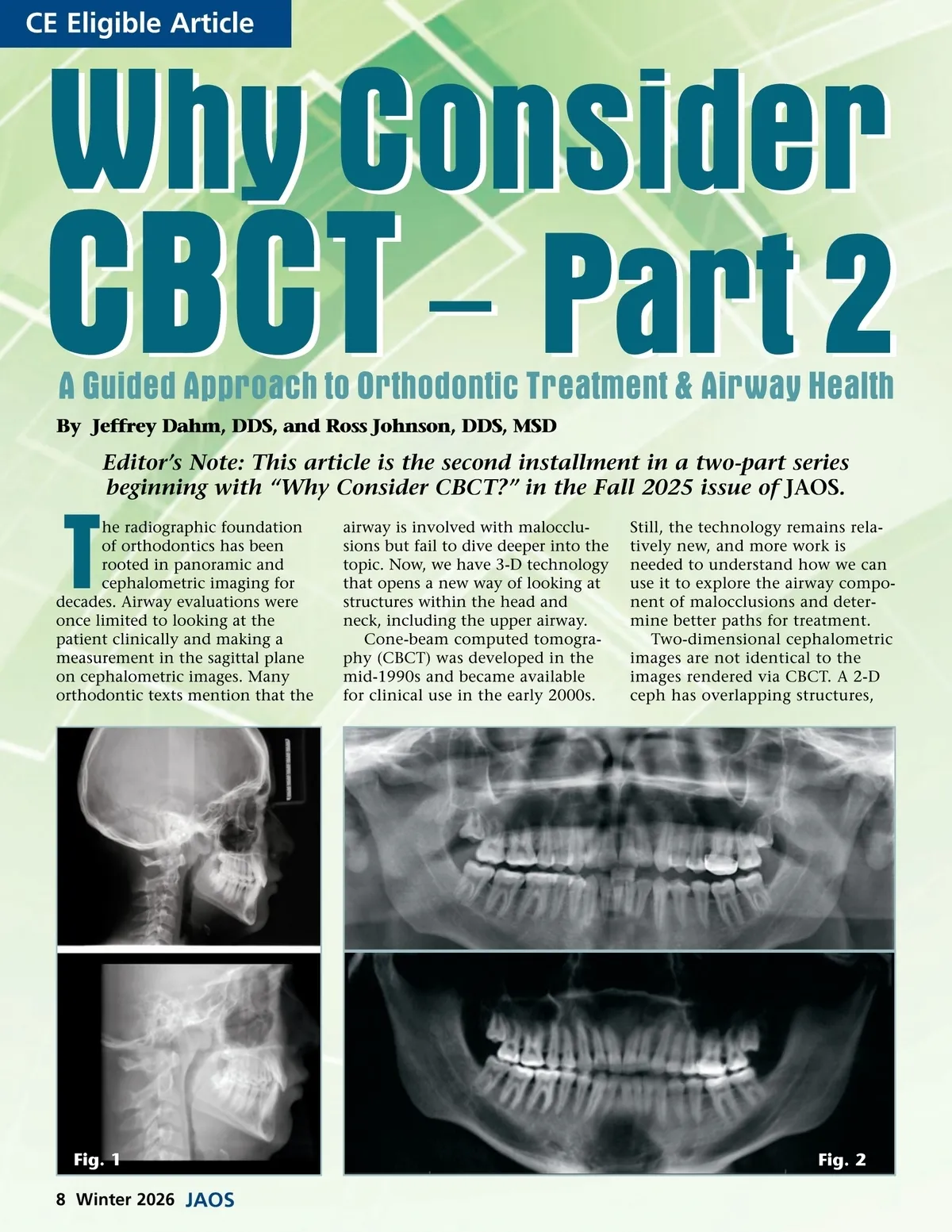
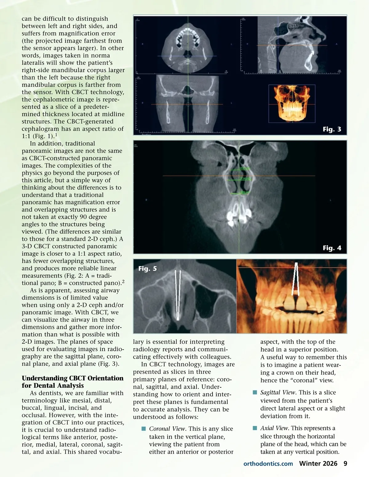
CE Eligible Article Why Consider CBCT – Part 2 A Guided Approach to Orthodontic Treatment & Airway Health By Jeffrey Dahm, DDS, and Ross Johnson, DDS, MSD Editor’s Note: This article is the second installment in a two-part series beginning with “Why Consider CBCT?” in the Fall 2025 issue of JAOS . T he radiographic foundation of orthodontics has been rooted in panoramic and cephalometric imaging for decades. Airway evaluations were once limited to looking at the patient clinically and making a measurement in the sagittal plane on cephalometric images. Many orthodontic texts mention that the airway is involved with malocclu-sions but fail to dive deeper into the topic. Now, we have 3-D technology that opens a new way of looking at structures within the head and neck, including the upper airway. Cone-beam computed tomogra-phy (CBCT) was developed in the mid-1990s and became available for clinical use in the early 2000s. Still, the technology remains rela-tively new, and more work is needed to understand how we can use it to explore the airway compo-nent of malocclusions and deter-mine better paths for treatment. Two-dimensional cephalometric images are not identical to the images rendered via CBCT. A 2-D ceph has overlapping structures, Fig. 1 8 Winter 2026 JAOS Fig. 2
Journal of the American Orthodontic Society Winter 2026: Page 8