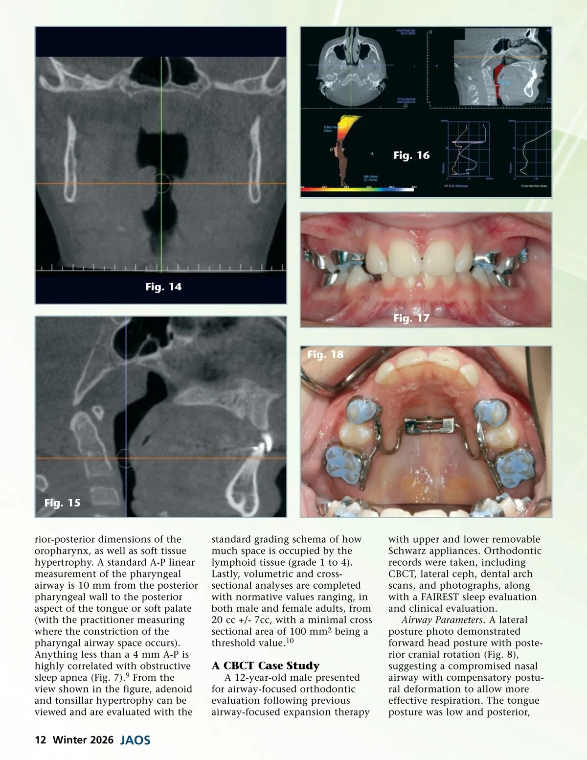
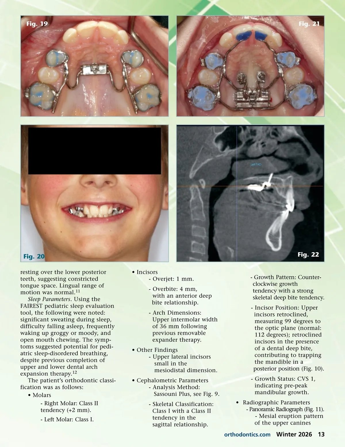
Fig. 16 Fig. 14 Fig. 17 Fig. 18 Fig. 15 rior-posterior dimensions of the oropharynx, as well as soft tissue hypertrophy. A standard A-P linear measurement of the pharyngeal airway is 10 mm from the posterior pharyngeal wall to the posterior aspect of the tongue or soft palate (with the practitioner measuring where the constriction of the pharyngal airway space occurs). Anything less than a 4 mm A-P is highly correlated with obstructive sleep apnea (Fig. 7). 9 From the view shown in the figure, adenoid and tonsillar hypertrophy can be viewed and are evaluated with the standard grading schema of how much space is occupied by the lymphoid tissue (grade 1 to 4). Lastly, volumetric and cross-sectional analyses are completed with normative values ranging, in both male and female adults, from 20 cc +/-7cc, with a minimal cross sectional area of 100 mm 2 being a threshold value. 10 A CBCT Case Study A 12-year-old male presented for airway-focused orthodontic evaluation following previous airway-focused expansion therapy with upper and lower removable Schwarz appliances. Orthodontic records were taken, including CBCT, lateral ceph, dental arch scans, and photographs, along with a FAIREST sleep evaluation and clinical evaluation. Airway Parameters . A lateral posture photo demonstrated forward head posture with poste-rior cranial rotation (Fig. 8), suggesting a compromised nasal airway with compensatory postu-ral deformation to allow more effective respiration. The tongue posture was low and posterior, 12 Winter 2026 JAOS
Journal of the American Orthodontic Society Winter 2026: Page 12