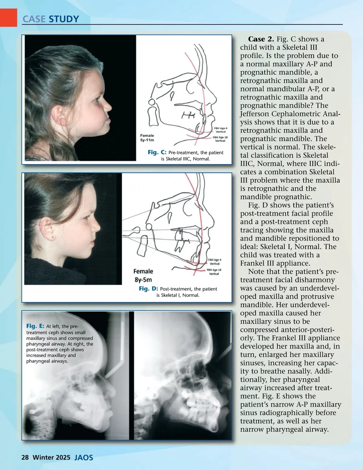
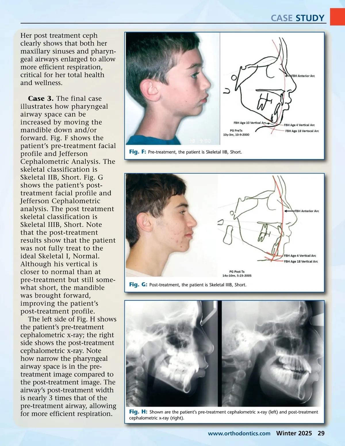
CASE STUDY Her post treatment ceph clearly shows that both her maxillary sinuses and pharyn-geal airways enlarged to allow more efficient respiration, critical for her total health and wellness. Case 3. The final case illustrates how pharyngeal airway space can be increased by moving the mandible down and/or forward. Fig. F shows the patient’s pre-treatment facial profile and Jefferson Cephalometric Analysis. The skeletal classification is Skeletal IIB, Short. Fig. G shows the patient’s post-treatment facial profile and Jefferson Cephalometric analysis. The post treatment skeletal classification is Skeletal IIIB, Short. Note that the post-treatment results show that the patient was not fully treat to the ideal Skeletal I, Normal. Although his vertical is closer to normal than at pre-treatment but still some-what short, the mandible was brought forward, improving the patient’s post-treatment profile. The left side of Fig. H shows the patient’s pre-treatment cephalometric x-ray; the right side shows the post-treatment cephalometric x-ray. Note how narrow the pharyngeal airway space is in the pre-treatment image compared to the post-treatment image. The airway’s post-treatment width is nearly 3 times that of the pre-treatment airway, allowing for more efficient respiration. Fig. F: Pre-treatment, the patient is Skeletal IIB, Short. Fig. G: Post-treatment, the patient is Skeletal IIIB, Short. Fig. H: Shown are the patient’s pre-treatment cephalometric x-ray (left) and post-treatment cephalometric x-ray (right). www.orthodontics.com Winter 2025 29
Journal of the American Orthodontic Society Winter 2025: Page 29