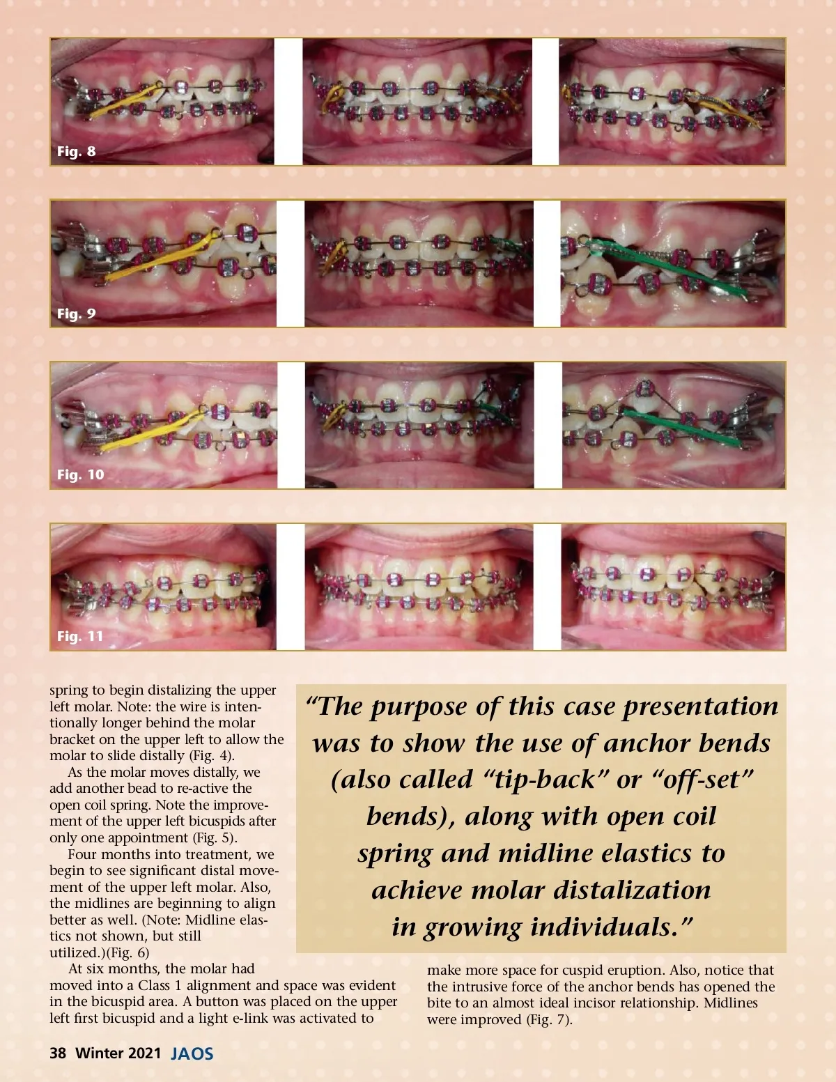
Fig. 8 Fig. 9 Fig. 10 Fig. 11 spring to begin distalizing the upper left molar. Note: the wire is inten-tionally longer behind the molar bracket on the upper left to allow the molar to slide distally (Fig. 4). As the molar moves distally, we add another bead to re-active the open coil spring. Note the improve-ment of the upper left bicuspids after only one appointment (Fig. 5). Four months into treatment, we begin to see significant distal move-ment of the upper left molar. Also, the midlines are beginning to align better as well. (Note: Midline elas-tics not shown, but still utilized.)(Fig. 6) At six months, the molar had moved into a Class 1 alignment and space was evident in the bicuspid area. A button was placed on the upper left first bicuspid and a light e-link was activated to “The purpose of this case presentation was to show the use of anchor bends (also called “tip-back” or “off-set” bends), along with open coil spring and midline elastics to achieve molar distalization in growing individuals.” make more space for cuspid eruption. Also, notice that the intrusive force of the anchor bends has opened the bite to an almost ideal incisor relationship. Midlines were improved (Fig. 7). 38 Winter 2021 JAOS
Journal of the American Orthodontic Society Winter 2021: Page 38