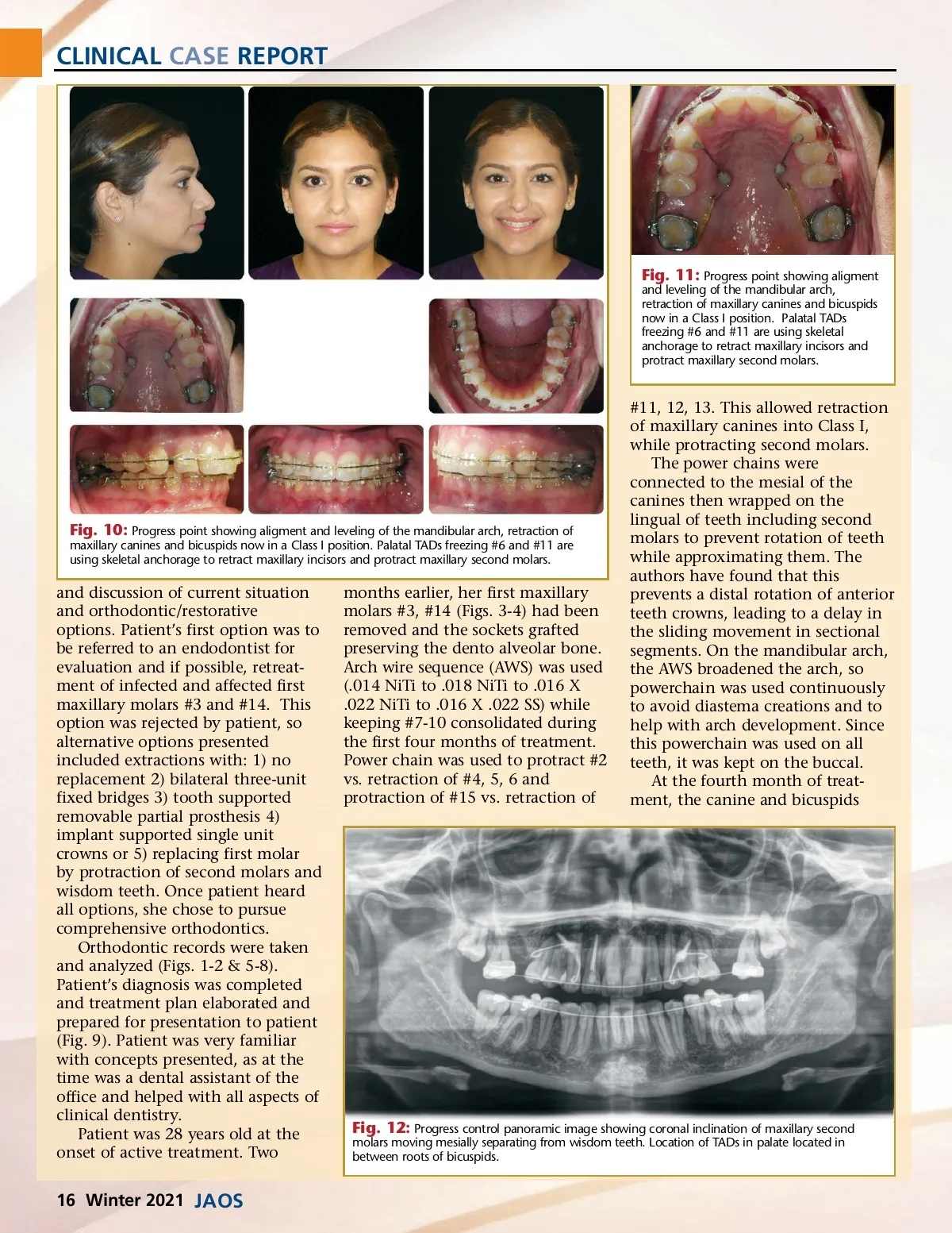
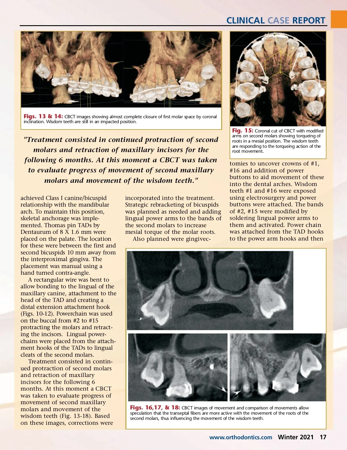
CLINICAL CASE REPORT Figs. 13 & 14: CBCT images showing almost complete closure of first molar space by coronal inclination. Wisdom teeth are still in an impacted position. "Treatment consisted in continued protraction of second molars and retraction of maxillary incisors for the following 6 months. At this moment a CBCT was taken to evaluate progress of movement of second maxillary molars and movement of the wisdom teeth." achieved Class I canine/bicuspid relationship with the mandibular arch. To maintain this position, skeletal anchorage was imple-mented. Thomas pin TADs by Dentaurum of 8 X 1.6 mm were placed on the palate. The location for these were between the first and second bicuspids 10 mm away from the interproximal gingiva. The placement was manual using a hand turned contra-angle. A rectangular wire was bent to allow bonding to the lingual of the maxillary canine, attachment to the head of the TAD and creating a distal extension attachment hook (Figs. 10-12). Powerchain was used on the buccal from #2 to #15 protracting the molars and retract-ing the incisors. Lingual power-chains were placed from the attach-ment hooks of the TADs to lingual cleats of the second molars. Treatment consisted in contin-ued protraction of second molars and retraction of maxillary incisors for the following 6 months. At this moment a CBCT was taken to evaluate progress of movement of second maxillary molars and movement of the wisdom teeth (Fig. 13-18). Based on these images, corrections were incorporated into the treatment. Strategic rebracketing of bicuspids was planned as needed and adding lingual power arms to the bands of the second molars to increase mesial torque of the molar roots. Also planned were gingivec-Fig. 15: Coronal cut of CBCT with modified arms on second molars showing torqueing of roots in a mesial position. The wisdom teeth are responding to the torqueing action of the root movement. tomies to uncover crowns of #1, #16 and addition of power buttons to aid movement of these into the dental arches. Wisdom teeth #1 and #16 were exposed using electrosurgery and power buttons were attached. The bands of #2, #15 were modified by soldering lingual power arms to them and activated. Power chain was attached from the TAD hooks to the power arm hooks and then Figs. 16,17, & 18: CBCT images of movement and comparison of movements allow speculation that the transeptal fibers are more active with the movement of the roots of the second molars, thus influencing the movement of the wisdom teeth. www.orthodontics.com Winter 2021 17
Journal of the American Orthodontic Society Winter 2021: Page 17