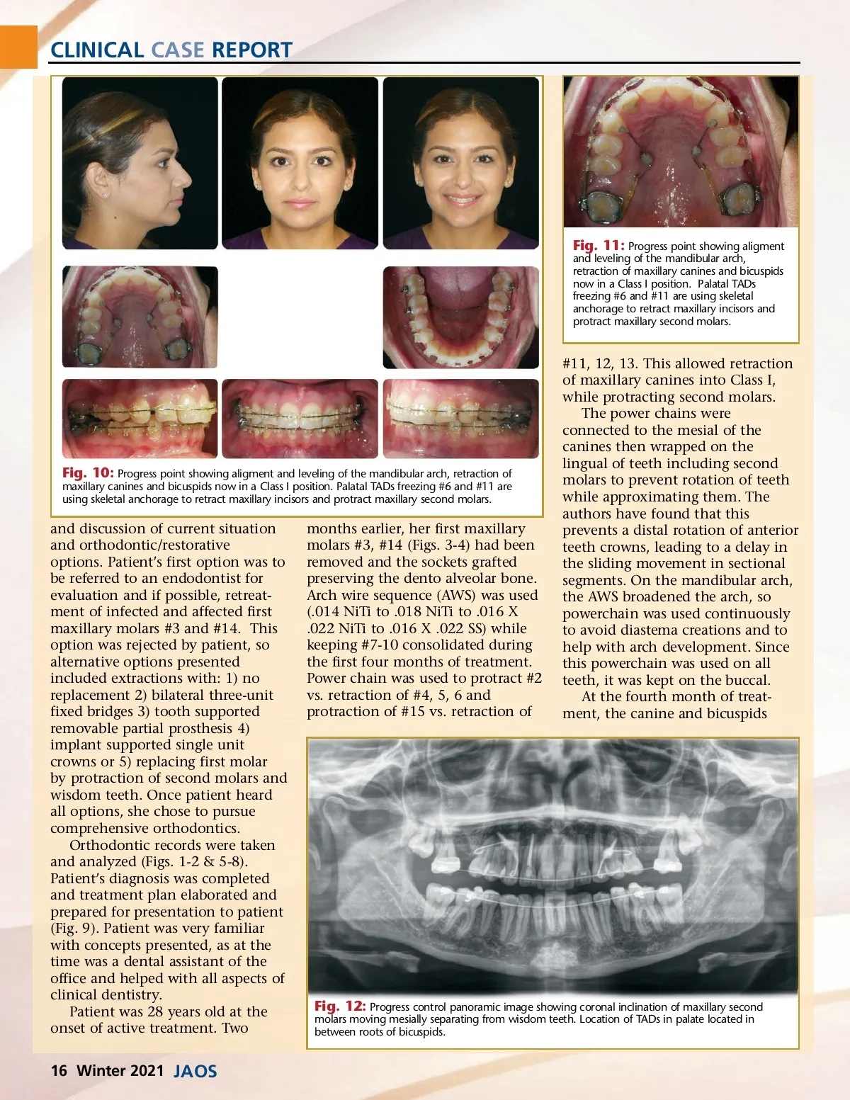
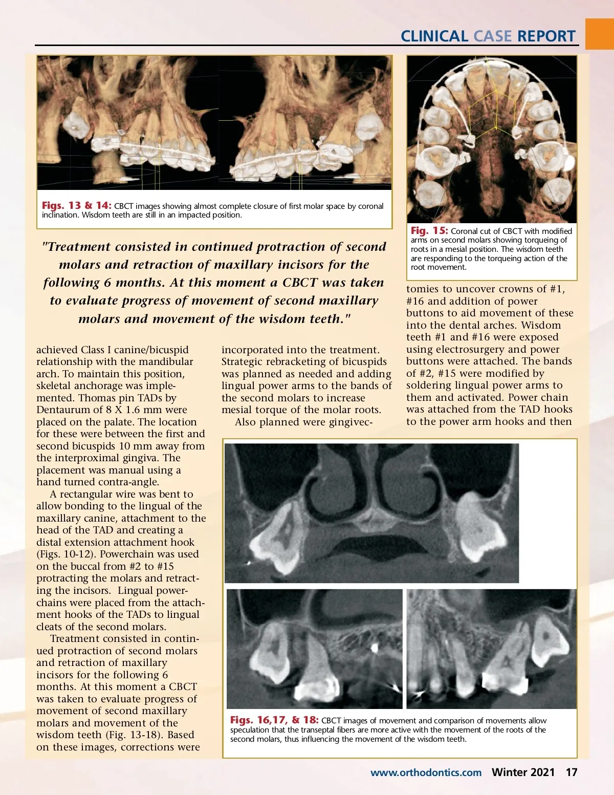
CLINICAL CASE REPORT Fig. 11: Progress point showing aligment and leveling of the mandibular arch, retraction of maxillary canines and bicuspids now in a Class I position. Palatal TADs freezing #6 and #11 are using skeletal anchorage to retract maxillary incisors and protract maxillary second molars. Fig. 10: Progress point showing aligment and leveling of the mandibular arch, retraction of maxillary canines and bicuspids now in a Class I position. Palatal TADs freezing #6 and #11 are using skeletal anchorage to retract maxillary incisors and protract maxillary second molars. and discussion of current situation and orthodontic/restorative options. Patient’s first option was to be referred to an endodontist for evaluation and if possible, retreat-ment of infected and affected first maxillary molars #3 and #14. This option was rejected by patient, so alternative options presented included extractions with: 1) no replacement 2) bilateral three-unit fixed bridges 3) tooth supported removable partial prosthesis 4) implant supported single unit crowns or 5) replacing first molar by protraction of second molars and wisdom teeth. Once patient heard all options, she chose to pursue comprehensive orthodontics. Orthodontic records were taken and analyzed (Figs. 1-2 & 5-8). Patient’s diagnosis was completed and treatment plan elaborated and prepared for presentation to patient (Fig. 9). Patient was very familiar with concepts presented, as at the time was a dental assistant of the office and helped with all aspects of clinical dentistry. Patient was 28 years old at the onset of active treatment. Two months earlier, her first maxillary molars #3, #14 (Figs. 3-4) had been removed and the sockets grafted preserving the dento alveolar bone. Arch wire sequence (AWS) was used (.014 NiTi to .018 NiTi to .016 X .022 NiTi to .016 X .022 SS) while keeping #7-10 consolidated during the first four months of treatment. Power chain was used to protract #2 vs. retraction of #4, 5, 6 and protraction of #15 vs. retraction of #11, 12, 13. This allowed retraction of maxillary canines into Class I, while protracting second molars. The power chains were connected to the mesial of the canines then wrapped on the lingual of teeth including second molars to prevent rotation of teeth while approximating them. The authors have found that this prevents a distal rotation of anterior teeth crowns, leading to a delay in the sliding movement in sectional segments. On the mandibular arch, the AWS broadened the arch, so powerchain was used continuously to avoid diastema creations and to help with arch development. Since this powerchain was used on all teeth, it was kept on the buccal. At the fourth month of treat-ment, the canine and bicuspids Fig. 12: Progress control panoramic image showing coronal inclination of maxillary second molars moving mesially separating from wisdom teeth. Location of TADs in palate located in between roots of bicuspids. 16 Winter 2021 JAOS
Journal of the American Orthodontic Society Winter 2021: Page 16