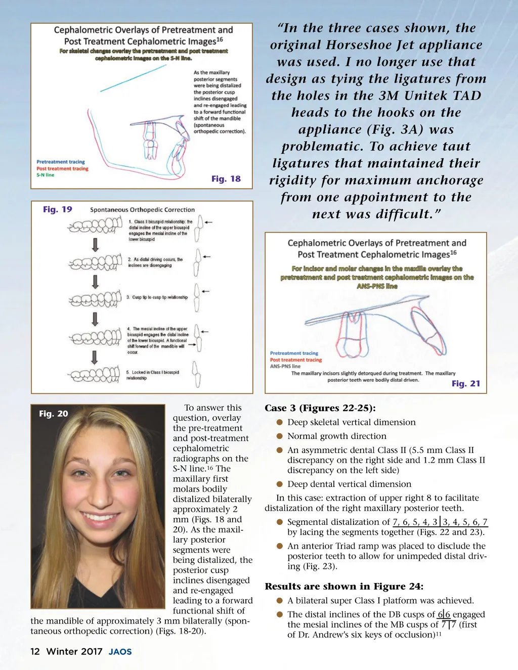
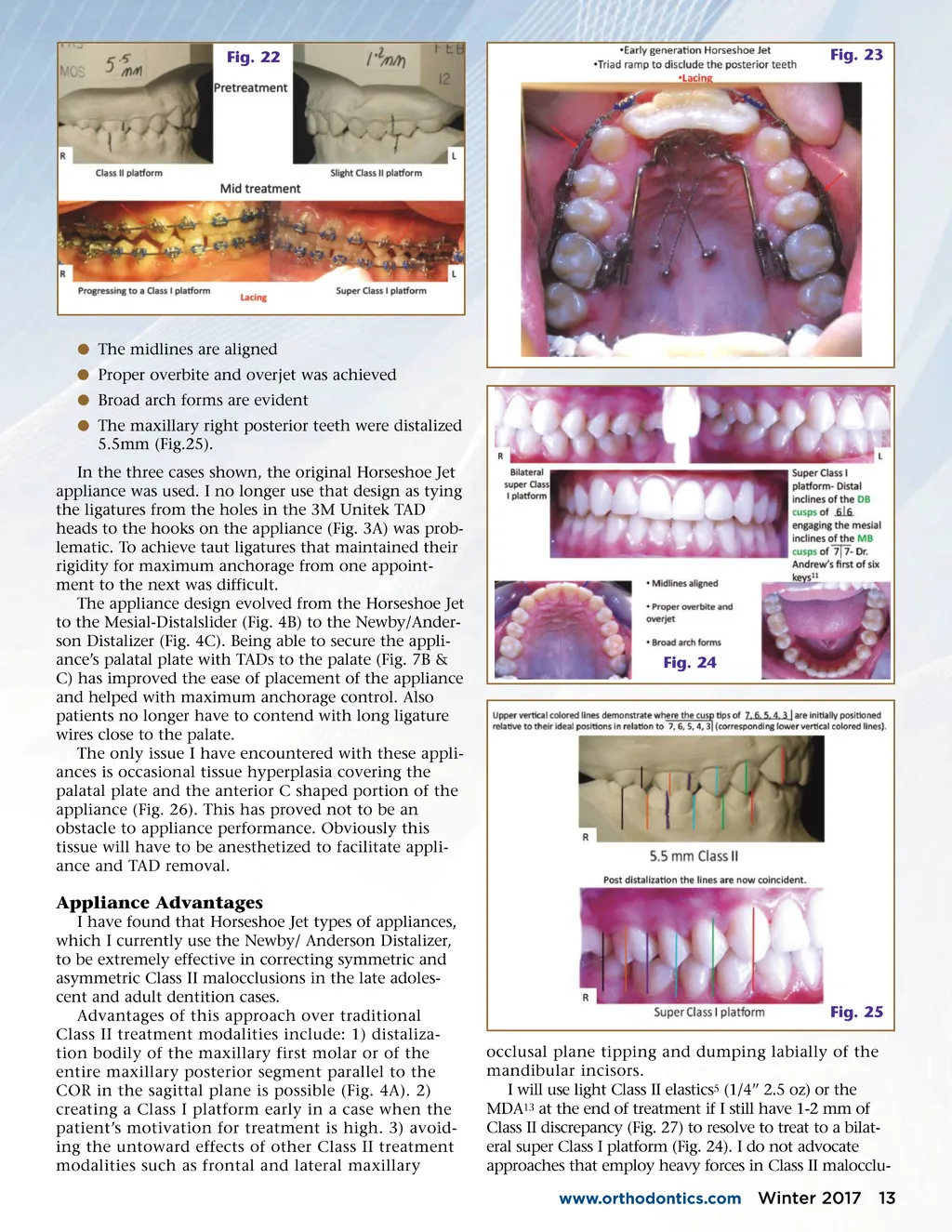
Fig. 18 Fig. 19 “In the three cases shown, the original Horseshoe Jet appliance was used. I no longer use that design as tying the ligatures from the holes in the 3M Unitek TAD heads to the hooks on the appliance (Fig. 3A) was problematic. To achieve taut ligatures that maintained their rigidity for maximum anchorage from one appointment to the next was difficult.” Fig. 21 To answer this question, overlay the pre-treatment and post-treatment cephalometric radiographs on the S-N line. 16 The maxillary first molars bodily distalized bilaterally approximately 2 mm (Figs. 18 and 20). As the maxil-lary posterior segments were being distalized, the posterior cusp inclines disengaged and re-engaged leading to a forward functional shift of the mandible of approximately 3 mm bilaterally (spon-taneous orthopedic correction) (Figs. 18-20). Fig. 20 Case 3 (Figures 22-25): b Deep skeletal vertical dimension b Normal growth direction b An asymmetric dental Class II (5.5 mm Class II discrepancy on the right side and 1.2 mm Class II discrepancy on the left side) b Deep dental vertical dimension In this case: extraction of upper right 8 to facilitate distalization of the right maxillary posterior teeth. b Segmental distalization of 7, 6, 5, 4, 3 3, 4, 5, 6, 7 by lacing the segments together (Figs. 22 and 23). b An anterior Triad ramp was placed to disclude the posterior teeth to allow for unimpeded distal driv-ing (Fig. 23). Results are shown in Figure 24: b A bilateral super Class I platform was achieved. b The distal inclines of the DB cusps of 6 6 engaged the mesial inclines of the MB cusps of 7 7 (first of Dr. Andrew’s six keys of occlusion) 11 12 Winter 2017 JAOS
Journal of the American Orthodontic Society Winter 2017: Page 12