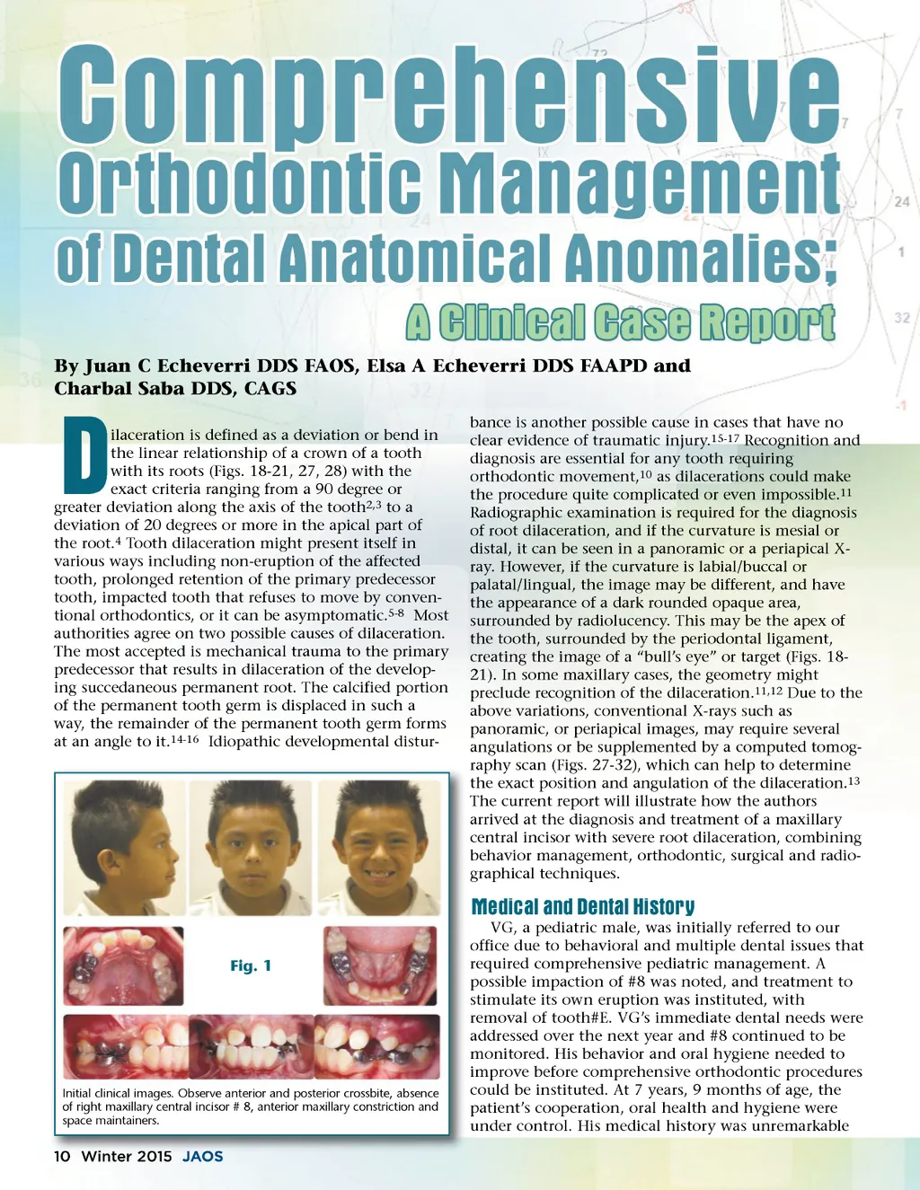
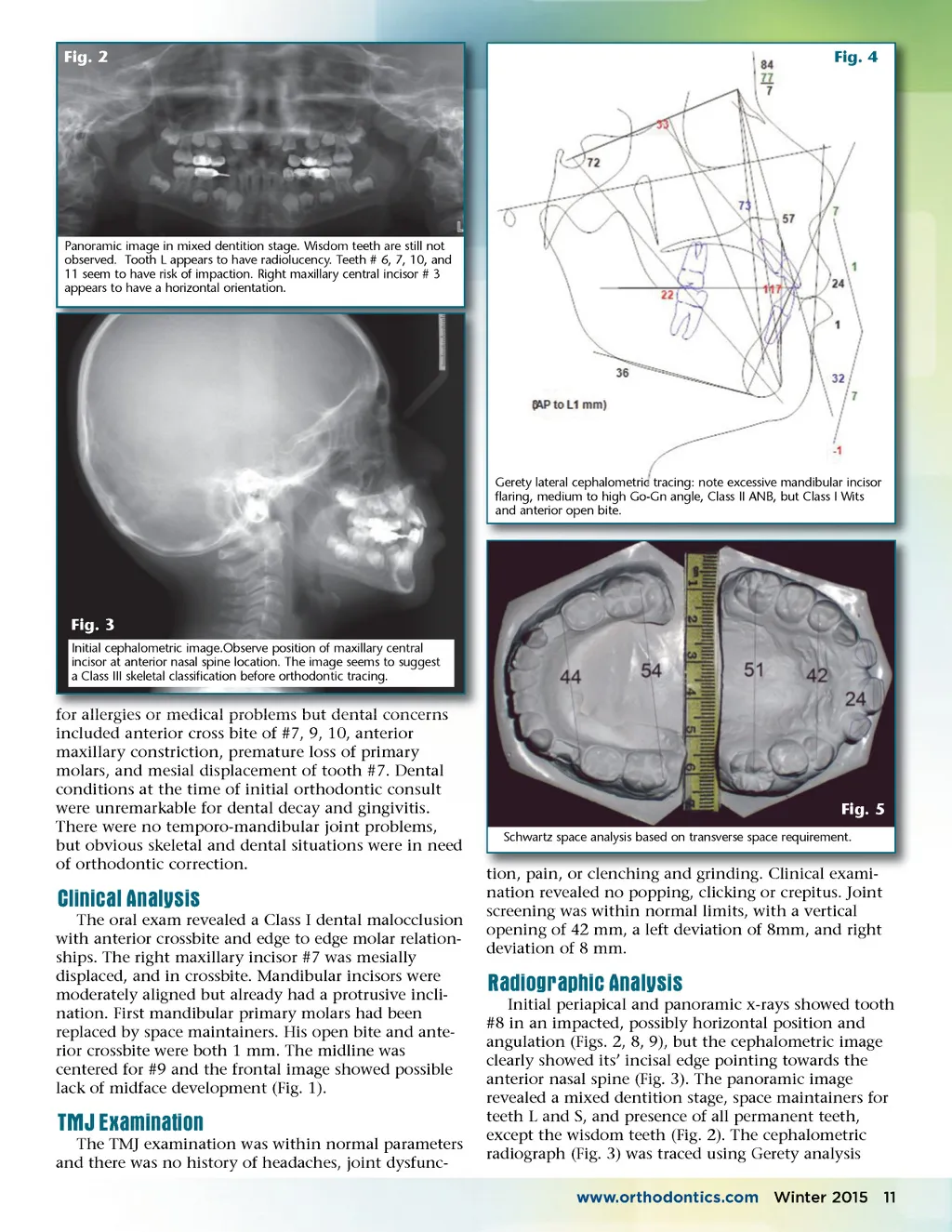
Fig. 2 Fig. 4 Panoramic image in mixed dentition stage. Wisdom teeth are still not observed. Tooth L appears to have radiolucency. Teeth # 6, 7, 10, and 11 seem to have risk of impaction. Right maxillary central incisor # 3 appears to have a horizontal orientation. Gerety lateral cephalometric tracing: note excessive mandibular incisor flaring, medium to high Go-Gn angle, Class II ANB, but Class I Wits and anterior open bite. Fig. 3 Initial cephalometric image.Observe position of maxillary central incisor at anterior nasal spine location. The image seems to suggest a Class III skeletal classification before orthodontic tracing. for allergies or medical problems but dental concerns included anterior cross bite of #7, 9, 10, anterior maxillary constriction, premature loss of primary molars, and mesial displacement of tooth #7. Dental conditions at the time of initial orthodontic consult were unremarkable for dental decay and gingivitis. There were no temporo-mandibular joint problems, but obvious skeletal and dental situations were in need of orthodontic correction. Fig. 5 Schwartz space analysis based on transverse space requirement. Clinical Analysis The oral exam revealed a Class I dental malocclusion with anterior crossbite and edge to edge molar relation-ships. The right maxillary incisor #7 was mesially displaced, and in crossbite. Mandibular incisors were moderately aligned but already had a protrusive incli-nation. First mandibular primary molars had been replaced by space maintainers. His open bite and ante-rior crossbite were both 1 mm. The midline was centered for #9 and the frontal image showed possible lack of midface development (Fig. 1). tion, tion pain, pain or clenching and grinding grinding. Clinical exami exami-nation revealed no popping, clicking or crepitus. Joint screening was within normal limits, with a vertical opening of 42 mm, a left deviation of 8mm, and right deviation of 8 mm. Radiographic Analysis Initial periapical and panoramic x-rays showed tooth #8 in an impacted, possibly horizontal position and angulation (Figs. 2, 8, 9), but the cephalometric image clearly showed its’ incisal edge pointing towards the anterior nasal spine (Fig. 3). The panoramic image revealed a mixed dentition stage, space maintainers for teeth L and S, and presence of all permanent teeth, except the wisdom teeth (Fig. 2). The cephalometric radiograph (Fig. 3) was traced using Gerety analysis www.orthodontics.com Winter 2015 11 TMJ Examination The TMJ examination was within normal parameters and there was no history of headaches, joint dysfunc-
Journal of the American Orthodontic Society Winter 2015: Page 11