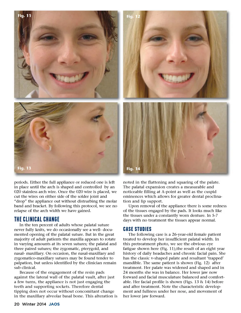
Fig. 11 Fig. 12 Fig. 13 Fig. 14 noted in the flattening and squaring of the palate. The palatal expansion creates a measurable and noticeable filling at A-point as well as the cuspid eminences which allows for greater dental proclina-tion and lip support. Upon removal of the appliance there is some redness of the tissues engaged by the pads. It looks much like the tissues under a constantly worn denture. In 5-7 days with no treatment the tissues appear normal. periods. Either the full appliance or reduced one is left in place until the arch is shaped and controlled by an 020 stainless arch wire. Once the 020 wire is placed, we cut the wires on either side of the solder joint and “drop” the appliance out without distrurbing the molar band and bracket. By following this protocol, we see no relapse of the arch width we have gained. '07&e;-.-&+e;7&+.0 In the ten percent of adults whose palatal suture never fully knits, we do occasionally see a well-docu-mented opening of the palatal suture. But in the great majority of adult patients the maxilla appears to rotate in varying amounts at its seven sutures; the palatal and three paired sutures; the zygomatic, pterygoid, and nasal-maxillary. On occasion, the nasal-maxillary and zygomatico-maxillary sutures may be found tender to palpation, but unless identified by the clinician remain sub clinical. Because of the engagement of the resin pads against the lateral wall of the palatal vault, after just a few turns, the appliance is not just engaging the teeth and supporting sockets. Therefore dental tipping does not occur without concomitant change in the maxillary alveolar basal bone. This alteration is 20 Winter 2014 JAOS (316715 )461 The following case is a 26-year-old female patient treated to develop her insufficient palatal width. In this pretreatment photo, we see the obvious eye fatigue shown here (Fig. 11),the result of an eight year history of daily headaches and chronic facial pain. She has the classic v-shaped palate and resultant ‘trapped’ mandible. The same patient is shown (Fig. 12) after treatment. Her palate was widened and shaped and in 24 months she was in balance. Her lower jaw now forward and facial musculature balanced and comfort-able. Her facial profile is shown (Figs. 13 & 14) before and after treatment. Note the characteristic develop-ment and fullness under her nose, and movement of her lower jaw forward.
Journal of the American Orthodontic Society Winter 2014: Page 20