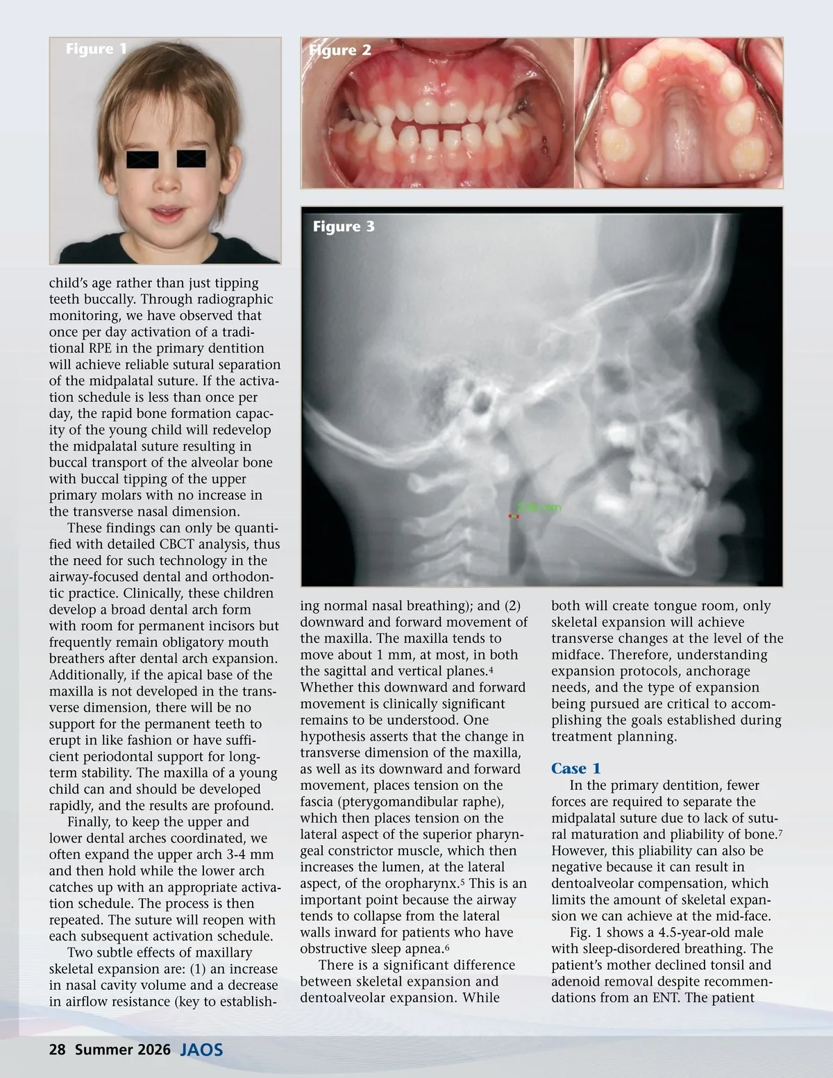
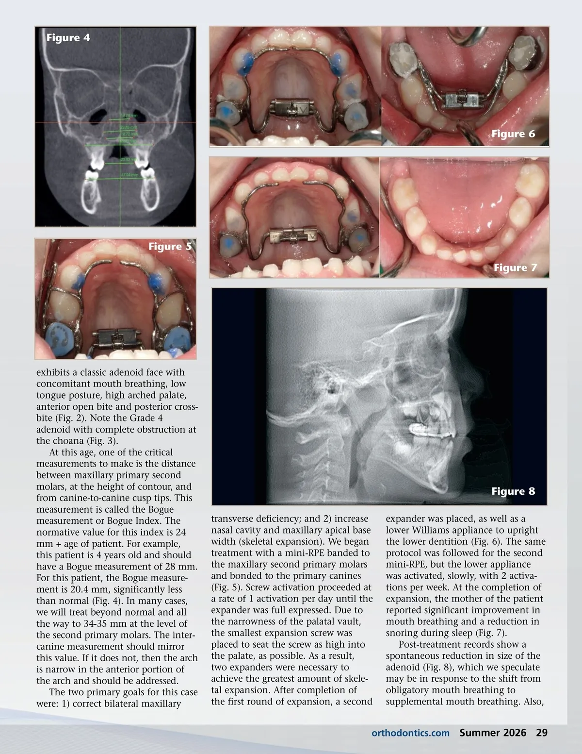
Figure 4 Figure 6 Figure 5 Figure 7 exhibits a classic adenoid face with concomitant mouth breathing, low tongue posture, high arched palate, anterior open bite and posterior cross-bite (Fig. 2). Note the Grade 4 adenoid with complete obstruction at the choana (Fig. 3). At this age, one of the critical measurements to make is the distance between maxillary primary second molars, at the height of contour, and from canine-to-canine cusp tips. This measurement is called the Bogue measurement or Bogue Index. The normative value for this index is 24 mm + age of patient. For example, this patient is 4 years old and should have a Bogue measurement of 28 mm. For this patient, the Bogue measure-ment is 20.4 mm, significantly less than normal (Fig. 4). In many cases, we will treat beyond normal and all the way to 34-35 mm at the level of the second primary molars. The inter-canine measurement should mirror this value. If it does not, then the arch is narrow in the anterior portion of the arch and should be addressed. The two primary goals for this case were: 1) correct bilateral maxillary Figure 8 transverse deficiency; and 2) increase nasal cavity and maxillary apical base width (skeletal expansion). We began treatment with a mini-RPE banded to the maxillary second primary molars and bonded to the primary canines (Fig. 5). Screw activation proceeded at a rate of 1 activation per day until the expander was full expressed. Due to the narrowness of the palatal vault, the smallest expansion screw was placed to seat the screw as high into the palate, as possible. As a result, two expanders were necessary to achieve the greatest amount of skele-tal expansion. After completion of the first round of expansion, a second expander was placed, as well as a lower Williams appliance to upright the lower dentition (Fig. 6). The same protocol was followed for the second mini-RPE, but the lower appliance was activated, slowly, with 2 activa-tions per week. At the completion of expansion, the mother of the patient reported significant improvement in mouth breathing and a reduction in snoring during sleep (Fig. 7). Post-treatment records show a spontaneous reduction in size of the adenoid (Fig. 8), which we speculate may be in response to the shift from obligatory mouth breathing to supplemental mouth breathing. Also, orthodontics.com Summer 2026 29
Journal of the American Orthodontic Society Summer 2026: Page 29