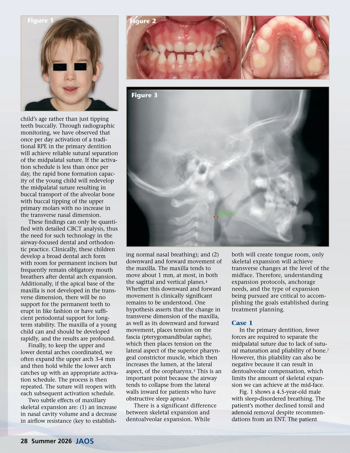
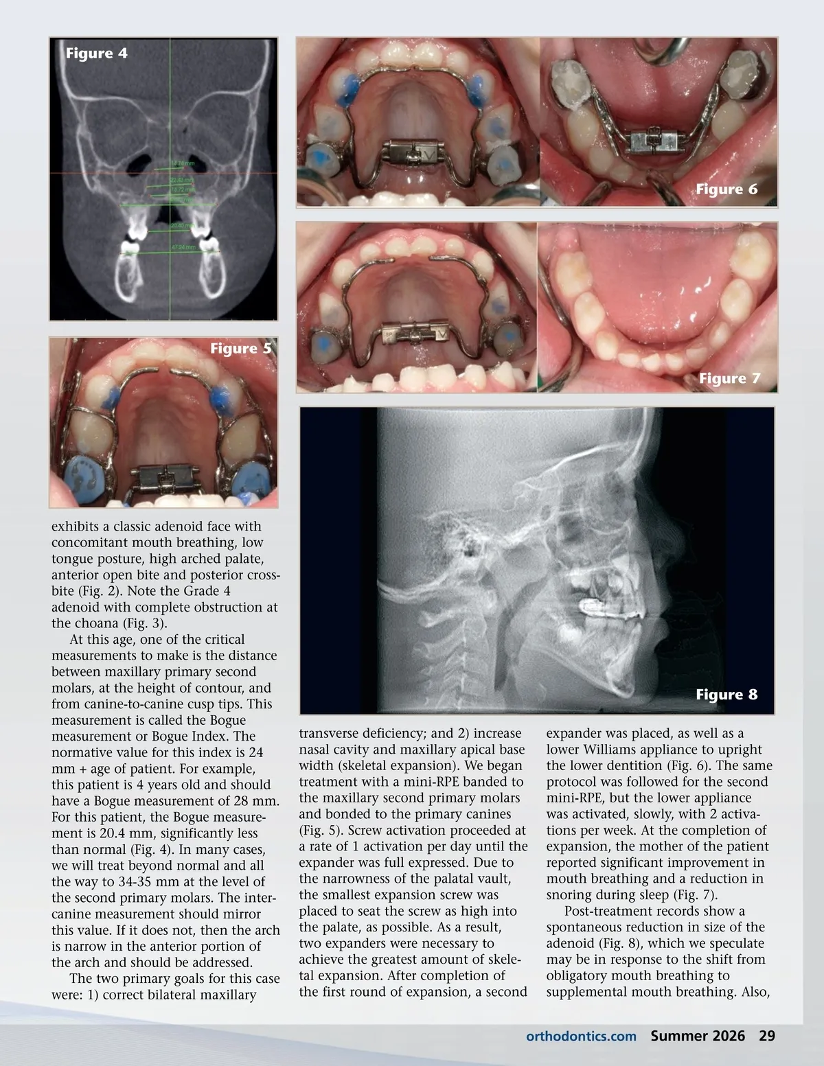
Figure 1 Figure 2 Figure 3 child’s age rather than just tipping teeth buccally. Through radiographic monitoring, we have observed that once per day activation of a tradi-tional RPE in the primary dentition will achieve reliable sutural separation of the midpalatal suture. If the activa-tion schedule is less than once per day, the rapid bone formation capac-ity of the young child will redevelop the midpalatal suture resulting in buccal transport of the alveolar bone with buccal tipping of the upper primary molars with no increase in the transverse nasal dimension. These findings can only be quanti-fied with detailed CBCT analysis, thus the need for such technology in the airway-focused dental and orthodon-tic practice. Clinically, these children develop a broad dental arch form with room for permanent incisors but frequently remain obligatory mouth breathers after dental arch expansion. Additionally, if the apical base of the maxilla is not developed in the trans-verse dimension, there will be no support for the permanent teeth to erupt in like fashion or have suffi-cient periodontal support for long-term stability. The maxilla of a young child can and should be developed rapidly, and the results are profound. Finally, to keep the upper and lower dental arches coordinated, we often expand the upper arch 3-4 mm and then hold while the lower arch catches up with an appropriate activa-tion schedule. The process is then repeated. The suture will reopen with each subsequent activation schedule. Two subtle effects of maxillary skeletal expansion are: (1) an increase in nasal cavity volume and a decrease in airflow resistance (key to establish-ing normal nasal breathing); and (2) downward and forward movement of the maxilla. The maxilla tends to move about 1 mm, at most, in both the sagittal and vertical planes. 4 Whether this downward and forward movement is clinically significant remains to be understood. One hypothesis asserts that the change in transverse dimension of the maxilla, as well as its downward and forward movement, places tension on the fascia (pterygomandibular raphe), which then places tension on the lateral aspect of the superior pharyn-geal constrictor muscle, which then increases the lumen, at the lateral aspect, of the oropharynx. 5 This is an important point because the airway tends to collapse from the lateral walls inward for patients who have obstructive sleep apnea. 6 There is a significant difference between skeletal expansion and dentoalveolar expansion. While both will create tongue room, only skeletal expansion will achieve transverse changes at the level of the midface. Therefore, understanding expansion protocols, anchorage needs, and the type of expansion being pursued are critical to accom-plishing the goals established during treatment planning. Case 1 In the primary dentition, fewer forces are required to separate the midpalatal suture due to lack of sutu-ral maturation and pliability of bone. 7 However, this pliability can also be negative because it can result in dentoalveolar compensation, which limits the amount of skeletal expan-sion we can achieve at the mid-face. Fig. 1 shows a 4.5-year-old male with sleep-disordered breathing. The patient’s mother declined tonsil and adenoid removal despite recommen-dations from an ENT. The patient 28 Summer 2026 JAOS
Journal of the American Orthodontic Society Summer 2026: Page 28