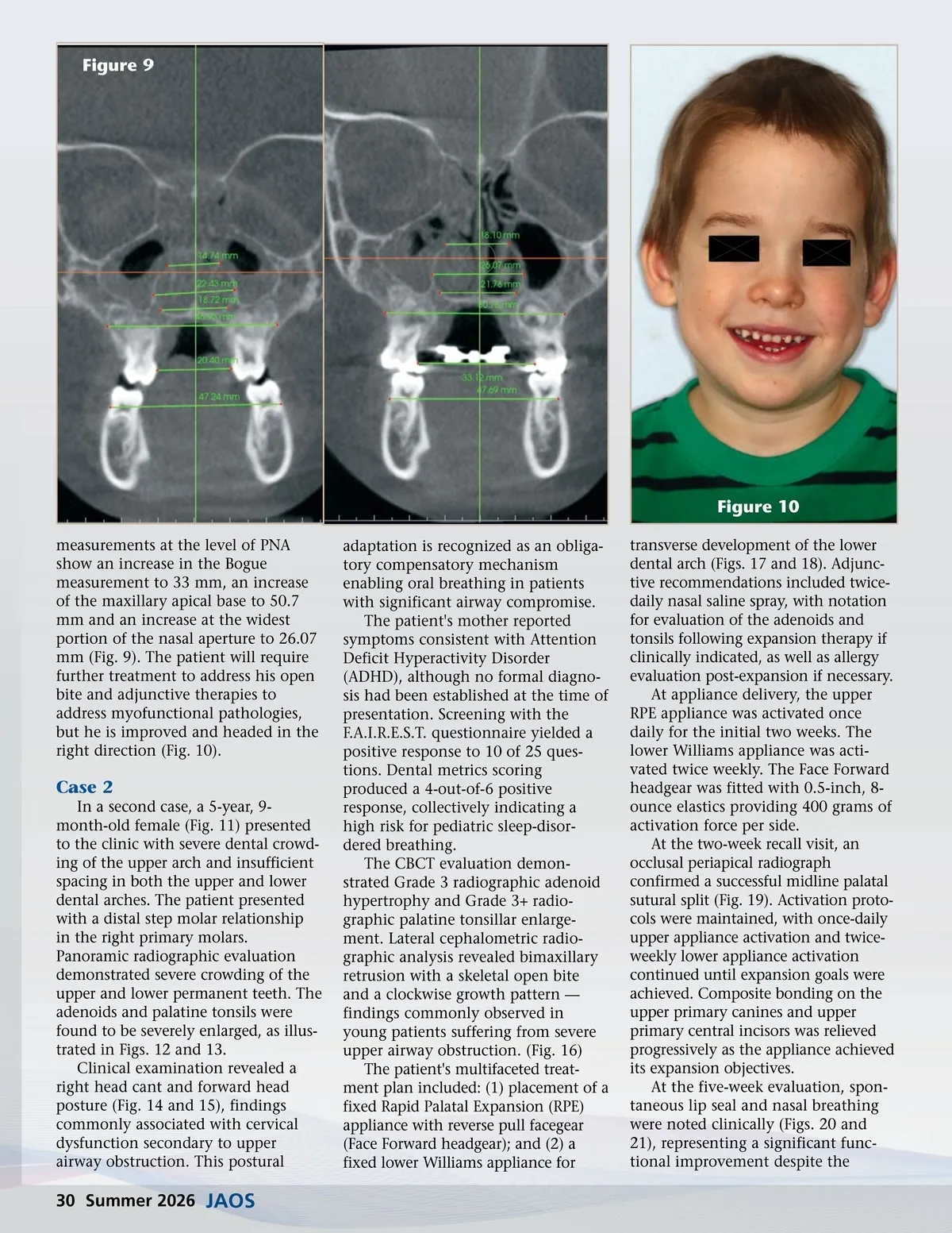
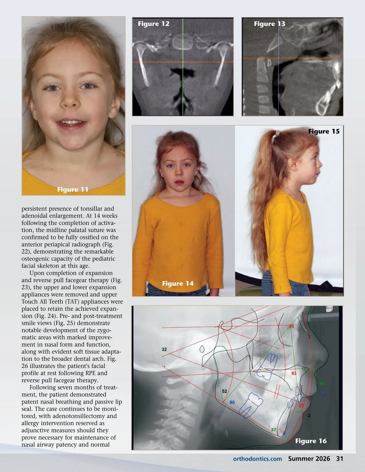
Figure 9 Figure 10 measurements at the level of PNA show an increase in the Bogue measurement to 33 mm, an increase of the maxillary apical base to 50.7 mm and an increase at the widest portion of the nasal aperture to 26.07 mm (Fig. 9). The patient will require further treatment to address his open bite and adjunctive therapies to address myofunctional pathologies, but he is improved and headed in the right direction (Fig. 10). adaptation is recognized as an obliga-tory compensatory mechanism enabling oral breathing in patients with significant airway compromise. The patient's mother reported symptoms consistent with Attention Deficit Hyperactivity Disorder (ADHD), although no formal diagno-sis had been established at the time of presentation. Screening with the F.A.I.R.E.S.T. questionnaire yielded a positive response to 10 of 25 ques-tions. Dental metrics scoring produced a 4-out-of-6 positive response, collectively indicating a high risk for pediatric sleep-disor-dered breathing. The CBCT evaluation demon-strated Grade 3 radiographic adenoid hypertrophy and Grade 3+ radio-graphic palatine tonsillar enlarge-ment. Lateral cephalometric radio-graphic analysis revealed bimaxillary retrusion with a skeletal open bite and a clockwise growth pattern — findings commonly observed in young patients suffering from severe upper airway obstruction. (Fig. 16) The patient's multifaceted treat-ment plan included: (1) placement of a fixed Rapid Palatal Expansion (RPE) appliance with reverse pull facegear (Face Forward headgear); and (2) a fixed lower Williams appliance for transverse development of the lower dental arch (Figs. 17 and 18). Adjunc-tive recommendations included twice-daily nasal saline spray, with notation for evaluation of the adenoids and tonsils following expansion therapy if clinically indicated, as well as allergy evaluation post-expansion if necessary. At appliance delivery, the upper RPE appliance was activated once daily for the initial two weeks. The lower Williams appliance was acti-vated twice weekly. The Face Forward headgear was fitted with 0.5-inch, 8-ounce elastics providing 400 grams of activation force per side. At the two-week recall visit, an occlusal periapical radiograph confirmed a successful midline palatal sutural split (Fig. 19). Activation proto-cols were maintained, with once-daily upper appliance activation and twice-weekly lower appliance activation continued until expansion goals were achieved. Composite bonding on the upper primary canines and upper primary central incisors was relieved progressively as the appliance achieved its expansion objectives. At the five-week evaluation, spon-taneous lip seal and nasal breathing were noted clinically (Figs. 20 and 21), representing a significant func-tional improvement despite the Case 2 In a second case, a 5-year, 9-month-old female (Fig. 11) presented to the clinic with severe dental crowd-ing of the upper arch and insufficient spacing in both the upper and lower dental arches. The patient presented with a distal step molar relationship in the right primary molars. Panoramic radiographic evaluation demonstrated severe crowding of the upper and lower permanent teeth. The adenoids and palatine tonsils were found to be severely enlarged, as illus-trated in Figs. 12 and 13. Clinical examination revealed a right head cant and forward head posture (Fig. 14 and 15), findings commonly associated with cervical dysfunction secondary to upper airway obstruction. This postural 30 Summer 2026 JAOS
Journal of the American Orthodontic Society Summer 2026: Page 30