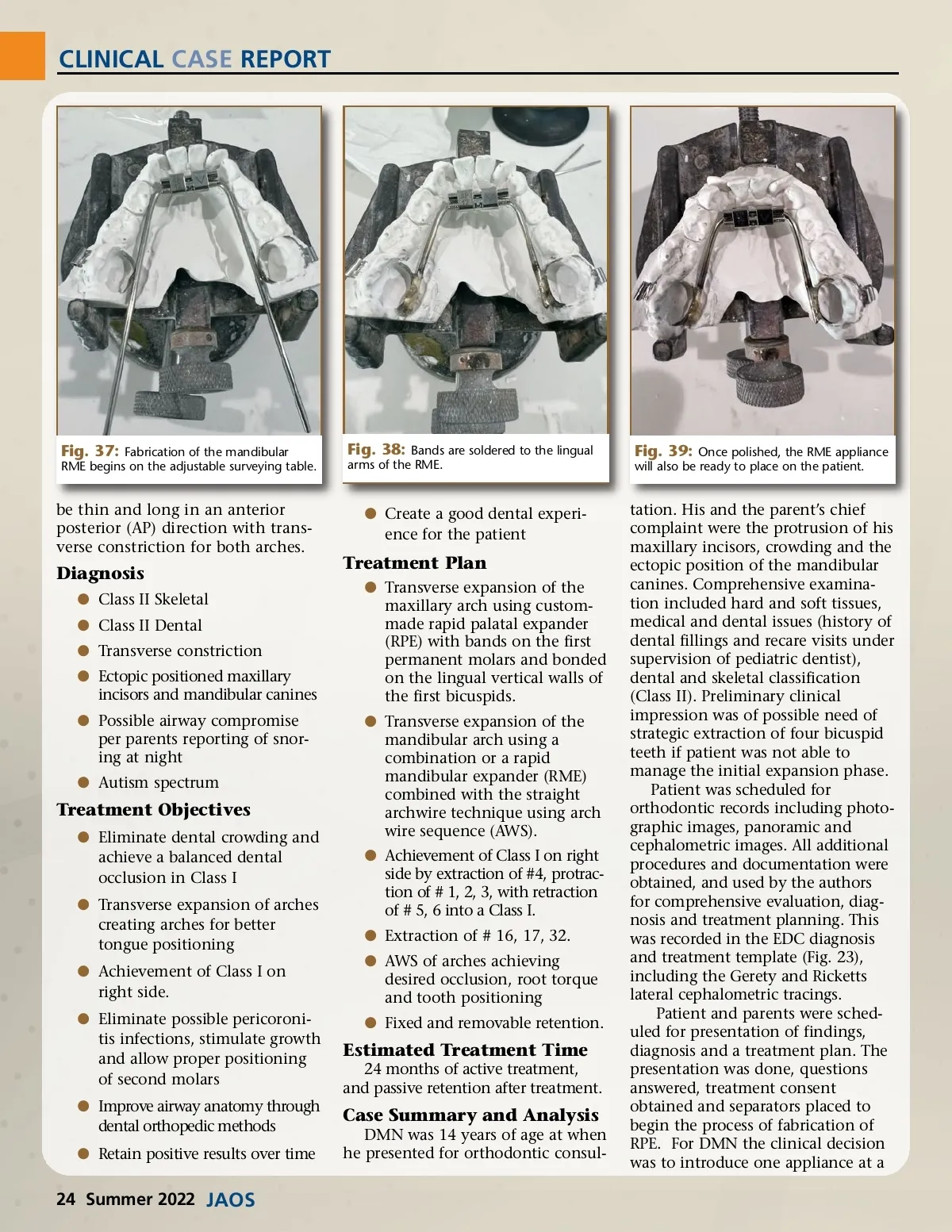
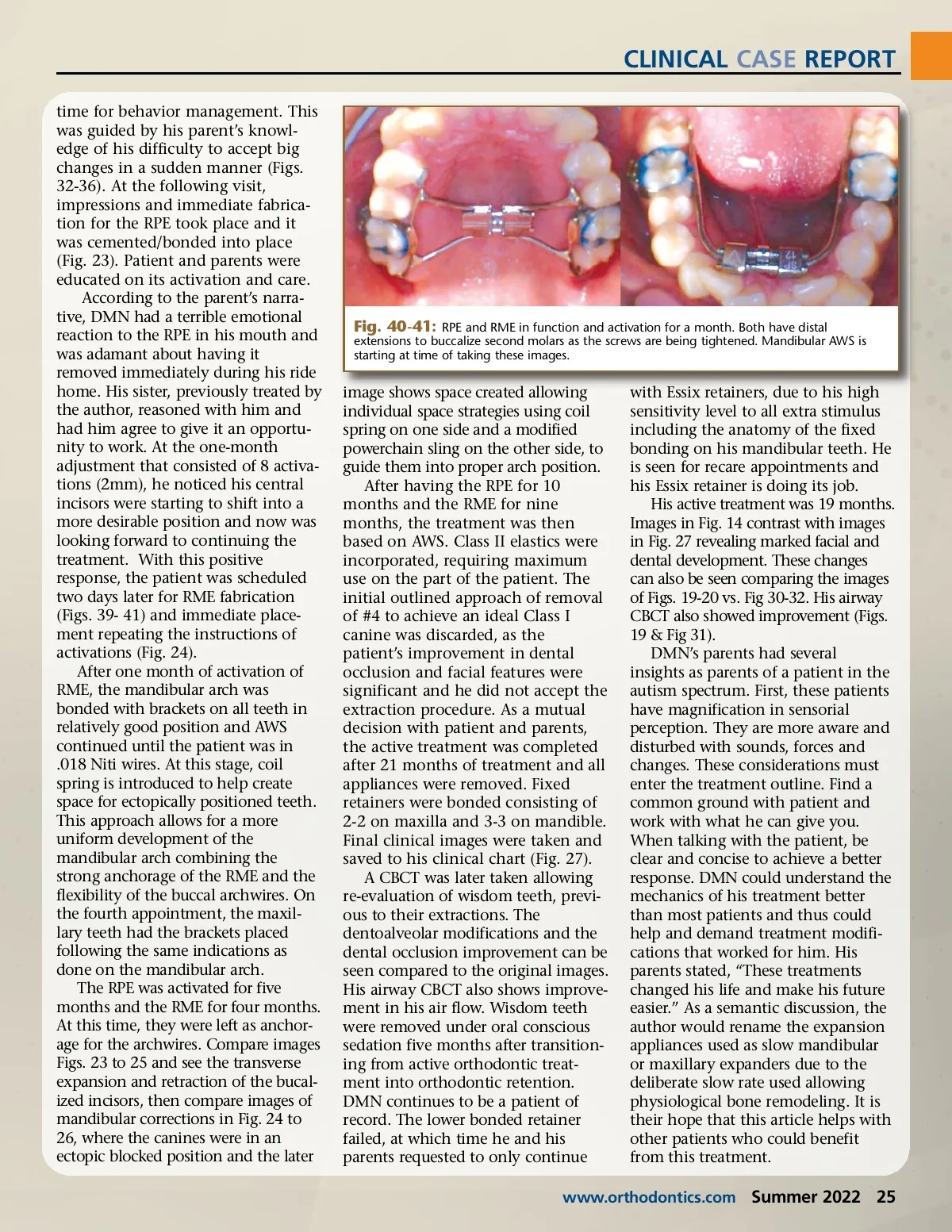
CLINICAL CASE REPORT time for behavior management. This was guided by his parent’s knowl-edge of his difficulty to accept big changes in a sudden manner (Figs. 32-36). At the following visit, impressions and immediate fabrica-tion for the RPE took place and it was cemented/bonded into place (Fig. 23). Patient and parents were educated on its activation and care. According to the parent’s narra-tive, DMN had a terrible emotional reaction to the RPE in his mouth and was adamant about having it removed immediately during his ride home. His sister, previously treated by the author, reasoned with him and had him agree to give it an opportu-nity to work. At the one-month adjustment that consisted of 8 activa-tions (2mm), he noticed his central incisors were starting to shift into a more desirable position and now was looking forward to continuing the treatment. With this positive response, the patient was scheduled two days later for RME fabrication (Figs. 39-41) and immediate place-ment repeating the instructions of activations (Fig. 24). After one month of activation of RME, the mandibular arch was bonded with brackets on all teeth in relatively good position and AWS continued until the patient was in .018 Niti wires. At this stage, coil spring is introduced to help create space for ectopically positioned teeth. This approach allows for a more uniform development of the mandibular arch combining the strong anchorage of the RME and the flexibility of the buccal archwires. On the fourth appointment, the maxil-lary teeth had the brackets placed following the same indications as done on the mandibular arch. The RPE was activated for five months and the RME for four months. At this time, they were left as anchor-age for the archwires. Compare images Figs. 23 to 25 and see the transverse expansion and retraction of the bucal-ized incisors, then compare images of mandibular corrections in Fig. 24 to 26, where the canines were in an ectopic blocked position and the later Fig. 40-41: RPE and RME in function and activation for a month. Both have distal extensions to buccalize second molars as the screws are being tightened. Mandibular AWS is starting at time of taking these images. image shows space created allowing individual space strategies using coil spring on one side and a modified powerchain sling on the other side, to guide them into proper arch position. After having the RPE for 10 months and the RME for nine months, the treatment was then based on AWS. Class II elastics were incorporated, requiring maximum use on the part of the patient. The initial outlined approach of removal of #4 to achieve an ideal Class I canine was discarded, as the patient’s improvement in dental occlusion and facial features were significant and he did not accept the extraction procedure. As a mutual decision with patient and parents, the active treatment was completed after 21 months of treatment and all appliances were removed. Fixed retainers were bonded consisting of 2-2 on maxilla and 3-3 on mandible. Final clinical images were taken and saved to his clinical chart (Fig. 27). A CBCT was later taken allowing re-evaluation of wisdom teeth, previ-ous to their extractions. The dentoalveolar modifications and the dental occlusion improvement can be seen compared to the original images. His airway CBCT also shows improve-ment in his air flow. Wisdom teeth were removed under oral conscious sedation five months after transition-ing from active orthodontic treat-ment into orthodontic retention. DMN continues to be a patient of record. The lower bonded retainer failed, at which time he and his parents requested to only continue with Essix retainers, due to his high sensitivity level to all extra stimulus including the anatomy of the fixed bonding on his mandibular teeth. He is seen for recare appointments and his Essix retainer is doing its job. His active treatment was 19 months. Images in Fig. 14 contrast with images in Fig. 27 revealing marked facial and dental development. These changes can also be seen comparing the images of Figs. 19-20 vs. Fig 30-32. His airway CBCT also showed improvement (Figs. 19 & Fig 31). DMN’s parents had several insights as parents of a patient in the autism spectrum. First, these patients have magnification in sensorial perception. They are more aware and disturbed with sounds, forces and changes. These considerations must enter the treatment outline. Find a common ground with patient and work with what he can give you. When talking with the patient, be clear and concise to achieve a better response. DMN could understand the mechanics of his treatment better than most patients and thus could help and demand treatment modifi-cations that worked for him. His parents stated, “These treatments changed his life and make his future easier.” As a semantic discussion, the author would rename the expansion appliances used as slow mandibular or maxillary expanders due to the deliberate slow rate used allowing physiological bone remodeling. It is their hope that this article helps with other patients who could benefit from this treatment. www.orthodontics.com Summer 2022 25
Journal of the American Orthodontic Society Summer 2022: Page 25