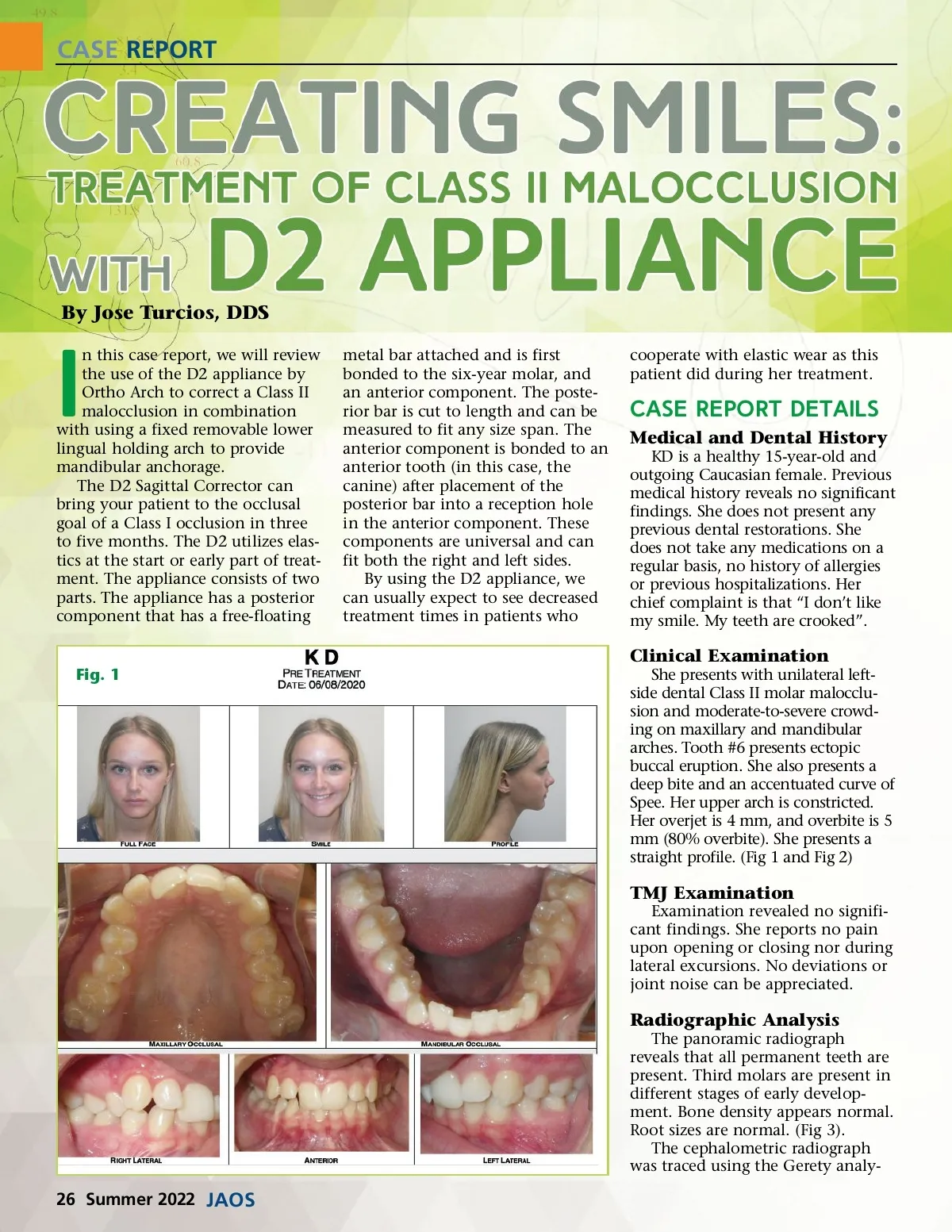
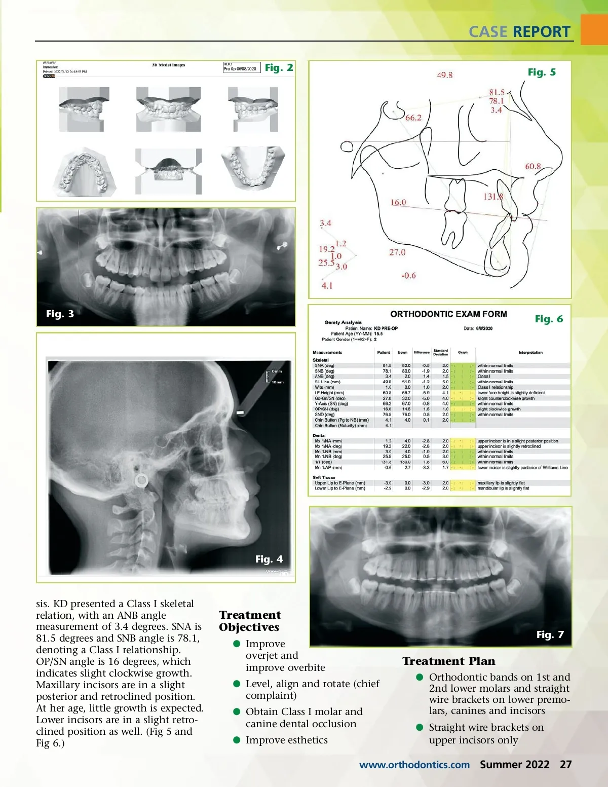
CASE REPORT By Jose Turcios, DDS I n this case report, we will review the use of the D2 appliance by Ortho Arch to correct a Class II malocclusion in combination with using a fixed removable lower lingual holding arch to provide mandibular anchorage. The D2 Sagittal Corrector can bring your patient to the occlusal goal of a Class I occlusion in three to five months. The D2 utilizes elas-tics at the start or early part of treat-ment. The appliance consists of two parts. The appliance has a posterior component that has a free-floating metal bar attached and is first bonded to the six-year molar, and an anterior component. The poste-rior bar is cut to length and can be measured to fit any size span. The anterior component is bonded to an anterior tooth (in this case, the canine) after placement of the posterior bar into a reception hole in the anterior component. These components are universal and can fit both the right and left sides. By using the D2 appliance, we can usually expect to see decreased treatment times in patients who cooperate with elastic wear as this patient did during her treatment. Case Report Details Medical and Dental History KD is a healthy 15-year-old and outgoing Caucasian female. Previous medical history reveals no significant findings. She does not present any previous dental restorations. She does not take any medications on a regular basis, no history of allergies or previous hospitalizations. Her chief complaint is that “I don’t like my smile. My teeth are crooked”. Clinical Examination Fig. 1 She presents with unilateral left-side dental Class II molar malocclu-sion and moderate-to-severe crowd-ing on maxillary and mandibular arches. Tooth #6 presents ectopic buccal eruption. She also presents a deep bite and an accentuated curve of Spee. Her upper arch is constricted. Her overjet is 4 mm, and overbite is 5 mm (80% overbite). She presents a straight profile. (Fig 1 and Fig 2) TMJ Examination Examination revealed no signifi-cant findings. She reports no pain upon opening or closing nor during lateral excursions. No deviations or joint noise can be appreciated. Radiographic Analysis The panoramic radiograph reveals that all permanent teeth are present. Third molars are present in different stages of early develop-ment. Bone density appears normal. Root sizes are normal. (Fig 3). The cephalometric radiograph was traced using the Gerety analy-26 Summer 2022 JAOS
Journal of the American Orthodontic Society Summer 2022: Page 26