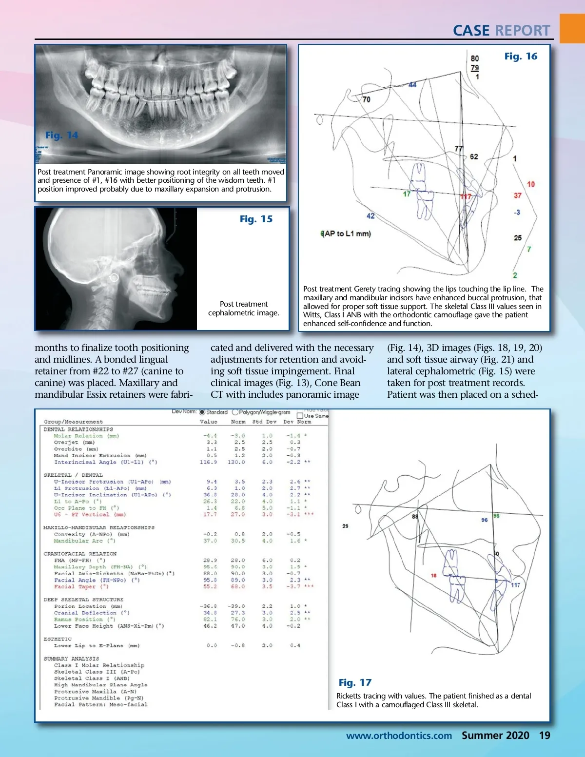
CASE REPORT Fig. 16 Fig. 14 Post treatment Panoramic image showing root integrity on all teeth moved and presence of #1, #16 with better positioning of the wisdom teeth. #1 position improved probably due to maxillary expansion and protrusion. Fig. 15 Post treatment cephalometric image. Post treatment Gerety tracing showing the lips touching the lip line. The maxillary and mandibular incisors have enhanced buccal protrusion, that allowed for proper soft tissue support. The skeletal Class III values seen in Witts, Class I ANB with the orthodontic camouflage gave the patient enhanced self-confidence and function. months to finalize tooth positioning and midlines. A bonded lingual retainer from #22 to #27 (canine to canine) was placed. Maxillary and mandibular Essix retainers were fabri-cated and delivered with the necessary adjustments for retention and avoid-ing soft tissue impingement. Final clinical images (Fig. 13), Cone Bean CT with includes panoramic image (Fig. 14), 3D images (Figs. 18, 19, 20) and soft tissue airway (Fig. 21) and lateral cephalometric (Fig. 15) were taken for post treatment records. Patient was then placed on a sched-Fig. 17 Ricketts tracing with values. The patient finished as a dental Class I with a camouflaged Class III skeletal. www.orthodontics.com Summer 2020 19
Journal of the American Orthodontic Society Summer 2020: Page 19