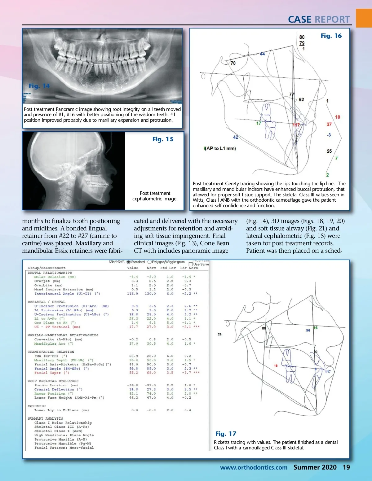
CASE REPORT Fig. 11 Fig. 12 Recare X-rays showing patient after removing RPE and D2 motion appliances and using straight wire appliances on the second phase of the orthodontic treatment. Treatment progress images after RPE expansion. The maxillar arch has an oval shape with elimination of crowding, increasing upper lip support and improving smile presentation. Mandibular teeth have canines out of ectopic position and incorporated into arch. mandible arch was treated using a D2 Motion appliance (Figs. 7, 8) as a bilat-eral mandibular tooth distalizer. For one-month elastics of 6.5 oz of force were used. After the fourth week, the elastics were increased to 8.5 oz. The mechanics behind this set-up were to achieve distalization of the mandibu-lar molars and bicuspids and protrac-tion of the maxillary arch while achieving lateral expansion. The patient was compliant and achieved good results (Figs. 9, 10). After nine weeks the patient was asked to stop activating the RPE, and all the maxil-lary teeth except the second molars had brackets with an .018 slot Roth prescription placed. The first bicuspids were released from their bonding to the RPE thus allowing their move-ment with the buccal archwire. The initial archwire was .014 NiTi to start alignment and leveling, correcting existing crowding and rounding the maxillary arch keeping the anchorage of the RPE. After four months of distalization, brackets were placed from #22 to #27 (mandibular canines and incisors) as a sectional correction. Class III elastics continued to be used distalizing the mandibular posterior teeth. The maxillary arch was advanced to an .018 NiTi archwire. Six weeks later the RPE and the D2 Motion appliances were removed simultaneously, brackets were placed on the mandibu-lar bicuspids and molars, and a .014 NiTi arch wire was placed to align and level. Light elastics with “short pull class III” were insti-tuted (#3 to #6 to #27/ #22 to #11 Fig. 13 to #14), creating a Class III vector that resisted open-ing the bite while stimulating Class III corrections by maxillary protru-Images of patient in retention. Patient has bonded lingual appliance sion and retention for maxillary incisors and for mandibula canines and incisors. mandibular Clear removable retainers are also used to enhance retention of the teeth. Patient’s facial features are better balanced and reports higher retraction. An levels of self-confidence. archwire sequence was followed as appliance vs. the cemented/bonded the teeth were moved into their new RPE. Gentle and slower movements positions (Figs. 11, 12). There were allowed the dento-alveolar bone time removable elastics used by patient and to remodel to the new dental posi-strategic powerchain by treating tions and adjustment of the patient’s doctor to complete movement of the neuro-muscular activities to her new teeth into a Class I dental occlusion. skeletal and dental anatomy. The For the next 11 months the patient maxillary arch received the lingual went through arch wire sequence bonded retainer from #7 to #10 progression and use of specific forces to retract or protract where needed to (lateral incisor to lateral incisor), and achieve and maintain the Class I posterior teeth allowed to settle into molar occlusion achieved by the occlusion. The mandibular arch distalization forces of the D2 motion continued being treated for two more 18 Summer 2020 JAOS
Journal of the American Orthodontic Society Summer 2020: Page 18