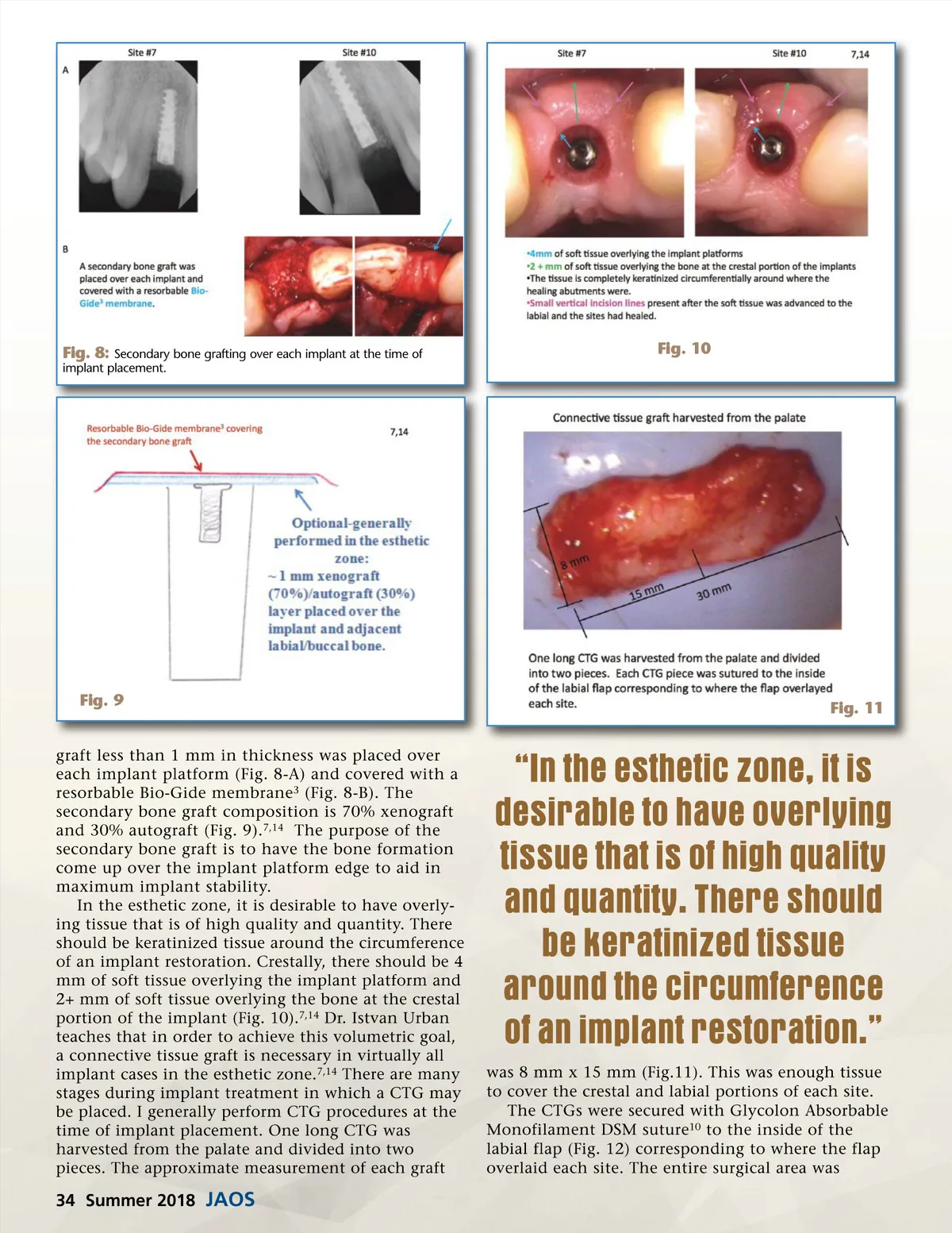
Fig. 12: Connective tissue grafting on the inside of the labial flap where the flap overlies each implant helps to increase tissue volume. Fig. 14 Fig. 13 sutured with GORE-TEX ePTFE Nonabsorbable Monofilament and primary closure was achieved (Fig. 13). Six months after implant placement and the CTG procedures were performed, the implants were uncovered. Small papilla sparing C-curve inci-sions were made a few millimeters lingual to each implant. Vertical connecting incisions were made on both sites of each implant on the labial (Fig. 14). These small flaps were advanced labially and all the tissue was preserved. The bone formation was so complete that the secondary bone graft entirely covered the platform of each implant (Fig. 15-A). A small trephine bur 9 was used to remove the bone overlying each implant without disturbing the bone at the implant edges. The cover screws were removed and NP 5x5 mm anatomic healing abutments 9 were placed (Fig. 15-B). The tissue flaps were advanced labially and rolled in against the labial surface of Fig. 15 each healing abutment and secured with Glycolon resorbable suture (Fig. 16). Six weeks were allowed for tissue maturation around the healing abutments. At the impression appointment, the healing abutments were removed and the tissue was inspected (Fig. 10). Note the tissue keratinization circumferentially around where the healing abutments were. Also note the 4+ mm of soft tissue height from the implant platforms to the soft tissue crests. There is an abundance of soft tissue (2+ mm) overlying the bone at the crestal portion of the implants. 7,14 Teeth 8 and 9 were prepped for conservative crowns due to stained and failing composite restora-www.orthodontics.com Summer 2018 35
Journal of the American Orthodontic Society Summer 2018/Buyer's Guide: Page 35