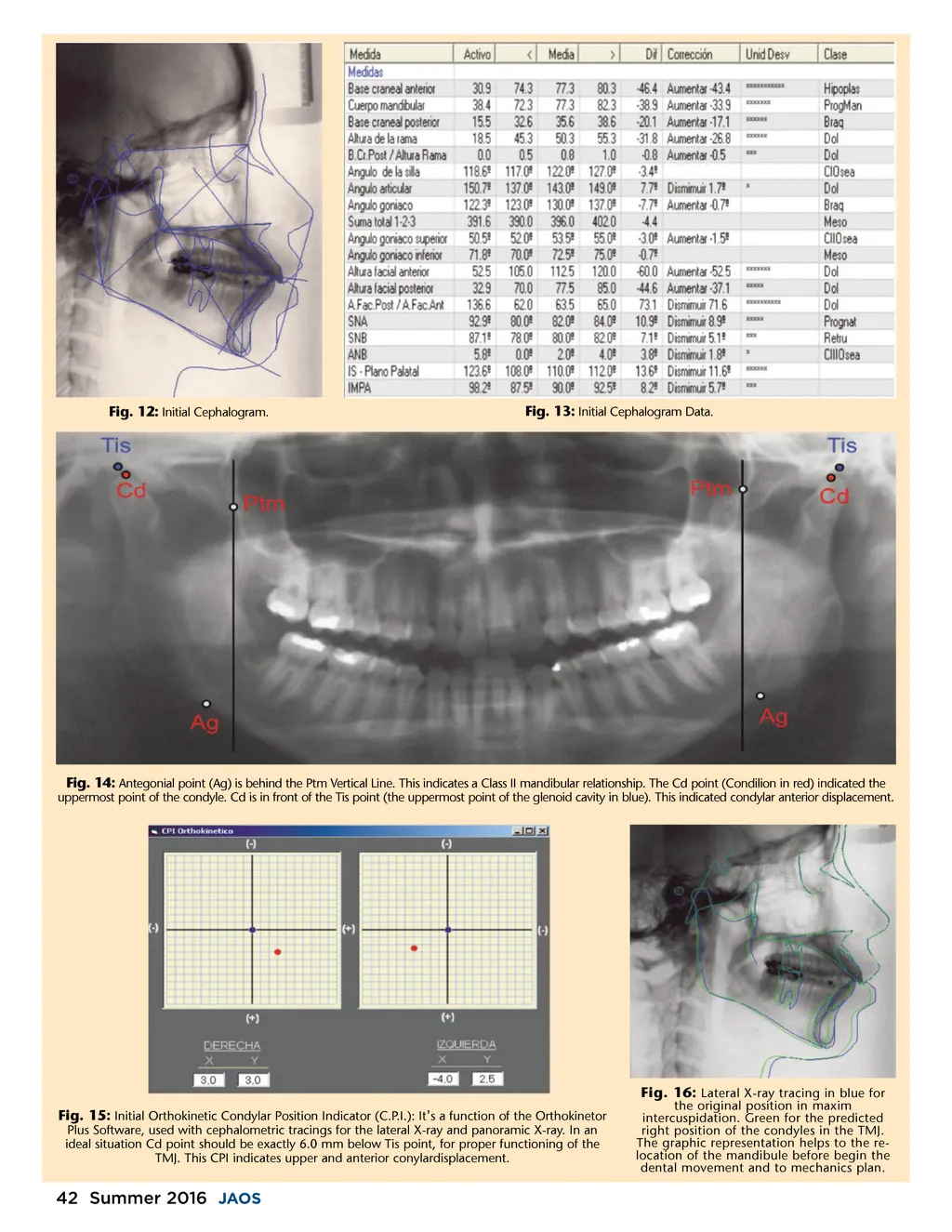
Fig. 12: Initial Cephalogram. Fig. 13: Initial Cephalogram Data. Fig. 14: Antegonial point (Ag) is behind the Ptm Vertical Line. This indicates a Class II mandibular relationship. The Cd point (Condilion in red) indicated the uppermost point of the condyle. Cd is in front of the Tis point (the uppermost point of the glenoid cavity in blue). This indicated condylar anterior displacement. Fig. 15: Initial Orthokinetic Condylar Position Indicator (C.P.I.): It ’ s a function of the Orthokinetor Plus Software, used with cephalometric tracings for the lateral X-ray and panoramic X-ray. In an ideal situation Cd point should be exactly 6.0 mm below Tis point, for proper functioning of the TMJ. This CPI indicates upper and anterior conylardisplacement. the original position in maxim intercuspidation. Green for the predicted right position of the condyles in the TMJ. The graphic representation helps to the re-location of the mandibule before begin the dental movement and to mechanics plan. Fig. 16: Lateral X-ray tracing in blue for 42 Summer 2016 JAOS
Journal of the American Orthodontic Society Summer 2016 / Buyer's Guide: Page 42