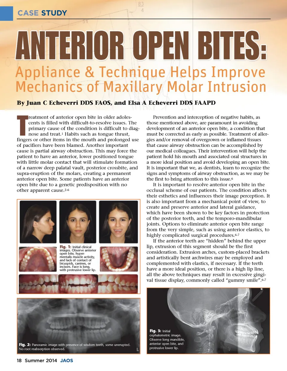
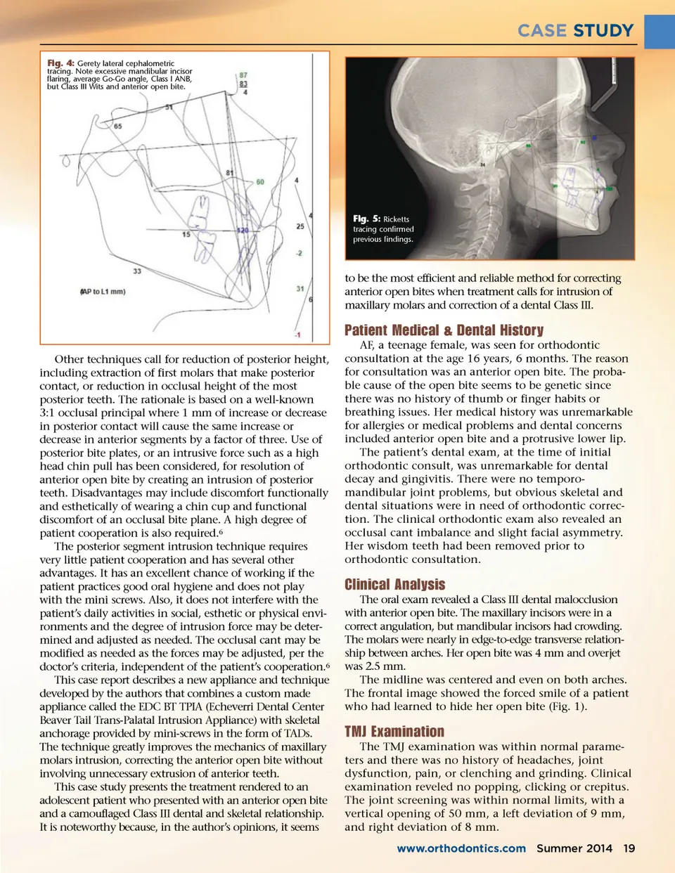
CASE STUDY Fig. 4: Gerety lateral cephalometric tracing. Note excessive mandibular incisor flaring, average Go-Go angle, Class I ANB, but Class III Wits and anterior open bite. Fig. 5: Ricketts tracing confirmed previous findings. to be the most efficient and reliable method for correcting anterior open bites when treatment calls for intrusion of maxillary molars and correction of a dental Class III. Patient Medical & Dental History Other techniques call for reduction of posterior height, including extraction of first molars that make posterior contact, or reduction in occlusal height of the most posterior teeth. The rationale is based on a well-known 3:1 occlusal principal where 1 mm of increase or decrease in posterior contact will cause the same increase or decrease in anterior segments by a factor of three. Use of posterior bite plates, or an intrusive force such as a high head chin pull has been considered, for resolution of anterior open bite by creating an intrusion of posterior teeth. Disadvantages may include discomfort functionally and esthetically of wearing a chin cup and functional discomfort of an occlusal bite plane. A high degree of patient cooperation is also required. 6 The posterior segment intrusion technique requires very little patient cooperation and has several other advantages. It has an excellent chance of working if the patient practices good oral hygiene and does not play with the mini screws. Also, it does not interfere with the patient’s daily activities in social, esthetic or physical envi-ronments and the degree of intrusion force may be deter-mined and adjusted as needed. The occlusal cant may be modified as needed as the forces may be adjusted, per the doctor’s criteria, independent of the patient’s cooperation. 6 This case report describes a new appliance and technique developed by the authors that combines a custom made appliance called the EDC BT TPIA (Echeverri Dental Center Beaver Tail Trans-Palatal Intrusion Appliance) with skeletal anchorage provided by mini-screws in the form of TADs. The technique greatly improves the mechanics of maxillary molars intrusion, correcting the anterior open bite without involving unnecessary extrusion of anterior teeth. This case study presents the treatment rendered to an adolescent patient who presented with an anterior open bite and a camouflaged Class III dental and skeletal relationship. It is noteworthy because, in the author’s opinions, it seems AF, a teenage female, was seen for orthodontic consultation at the age 16 years, 6 months. The reason for consultation was an anterior open bite. The proba-ble cause of the open bite seems to be genetic since there was no history of thumb or finger habits or breathing issues. Her medical history was unremarkable for allergies or medical problems and dental concerns included anterior open bite and a protrusive lower lip. The patient’s dental exam, at the time of initial orthodontic consult, was unremarkable for dental decay and gingivitis. There were no temporo-mandibular joint problems, but obvious skeletal and dental situations were in need of orthodontic correc-tion. The clinical orthodontic exam also revealed an occlusal cant imbalance and slight facial asymmetry. Her wisdom teeth had been removed prior to orthodontic consultation. Clinical Analysis The oral exam revealed a Class III dental malocclusion with anterior open bite. The maxillary incisors were in a correct angulation, but mandibular incisors had crowding. The molars were nearly in edge-to-edge transverse relation-ship between arches. Her open bite was 4 mm and overjet was 2.5 mm. The midline was centered and even on both arches. The frontal image showed the forced smile of a patient who had learned to hide her open bite (Fig. 1). TMJ Examination The TMJ examination was within normal parame-ters and there was no history of headaches, joint dysfunction, pain, or clenching and grinding. Clinical examination reveled no popping, clicking or crepitus. The joint screening was within normal limits, with a vertical opening of 50 mm, a left deviation of 9 mm, and right deviation of 8 mm. www.orthodontics.com Summer 2014 19
Journal of the American Orthodontic Society Summer 2014 / Buyer's Guide: Page 19