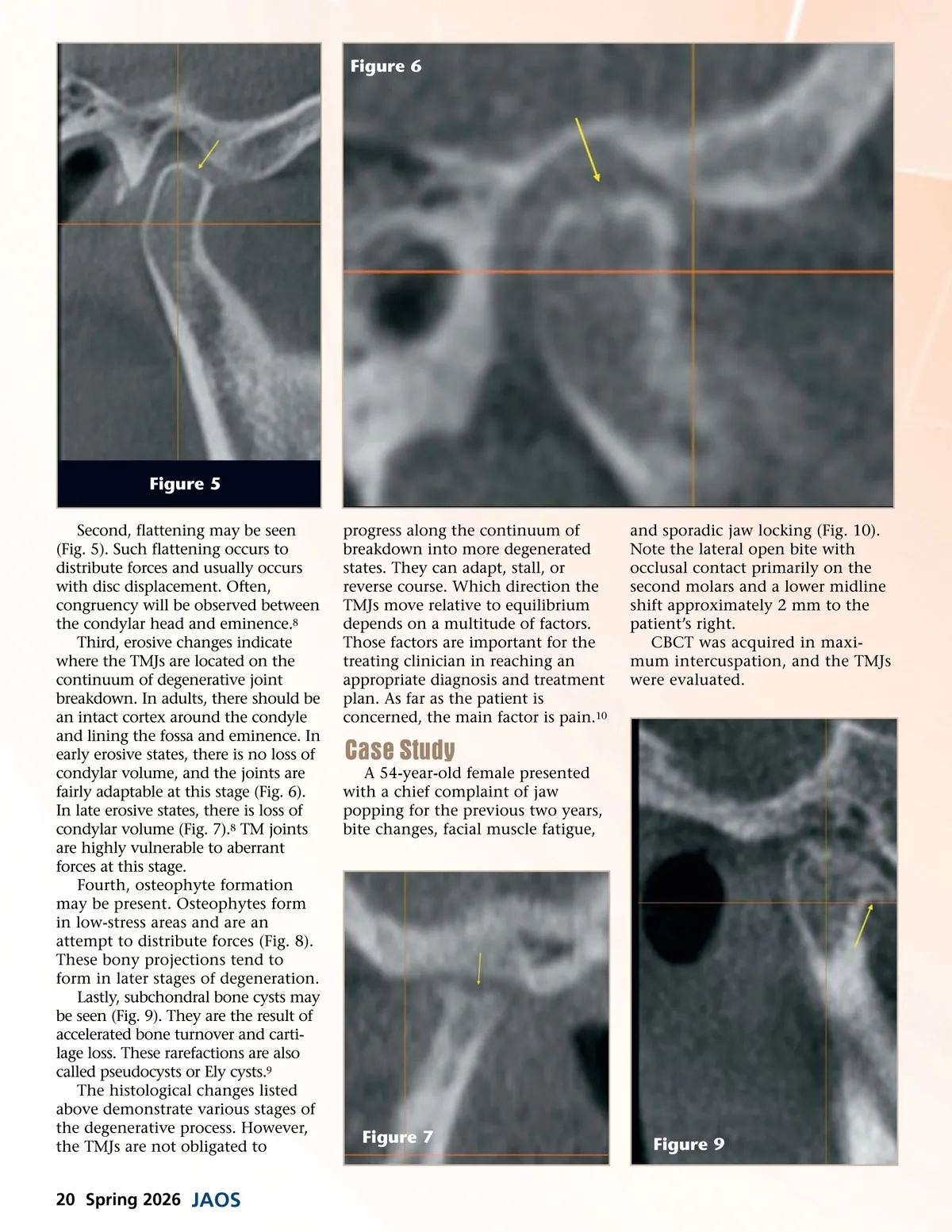
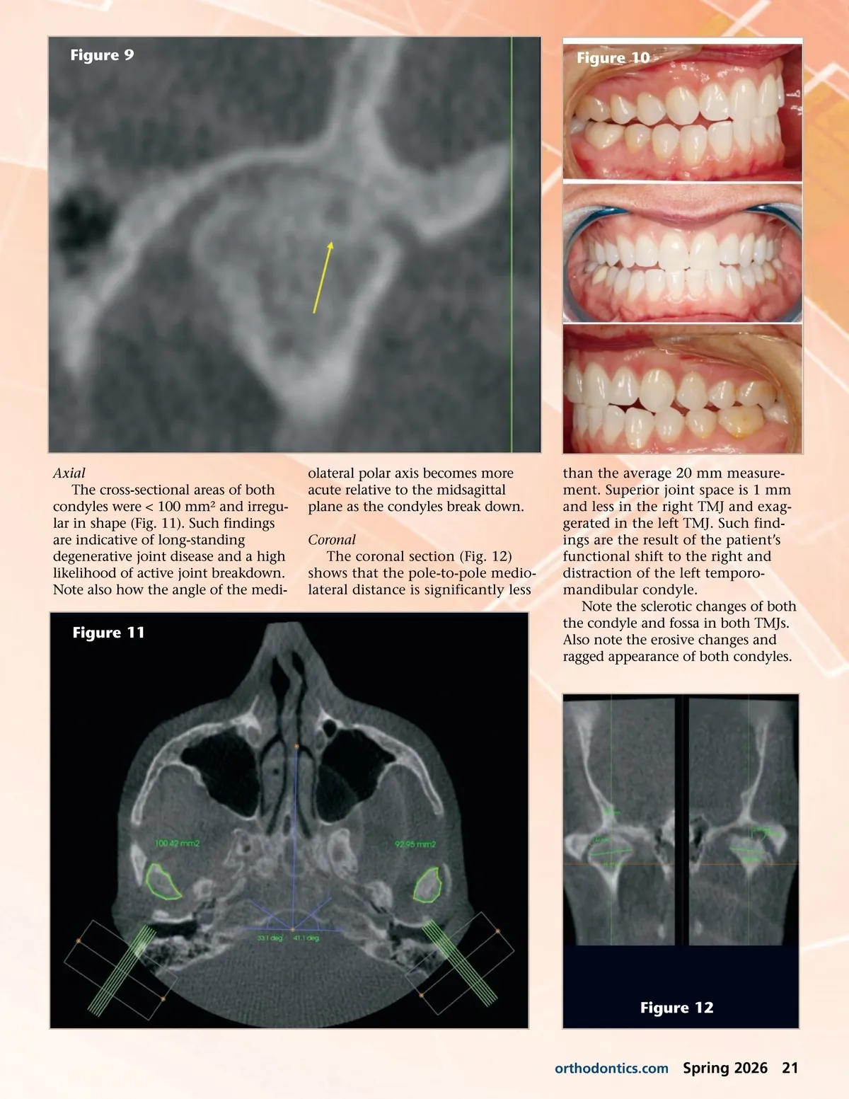
Figure 9 Figure 10 Axial The cross-sectional areas of both condyles were < 100 mm² and irregu-lar in shape (Fig. 11). Such findings are indicative of long-standing degenerative joint disease and a high likelihood of active joint breakdown. Note also how the angle of the medi-olateral polar axis becomes more acute relative to the midsagittal plane as the condyles break down. Coronal The coronal section (Fig. 12) shows that the pole-to-pole medio-lateral distance is significantly less Figure 11 than the average 20 mm measure-ment. Superior joint space is 1 mm and less in the right TMJ and exag-gerated in the left TMJ. Such find-ings are the result of the patient’s functional shift to the right and distraction of the left temporo-mandibular condyle. Note the sclerotic changes of both the condyle and fossa in both TMJs. Also note the erosive changes and ragged appearance of both condyles. Figure 12 orthodontics.com Spring 2026 21
Journal of the American Orthodontic Society Spring 2026: Page 21