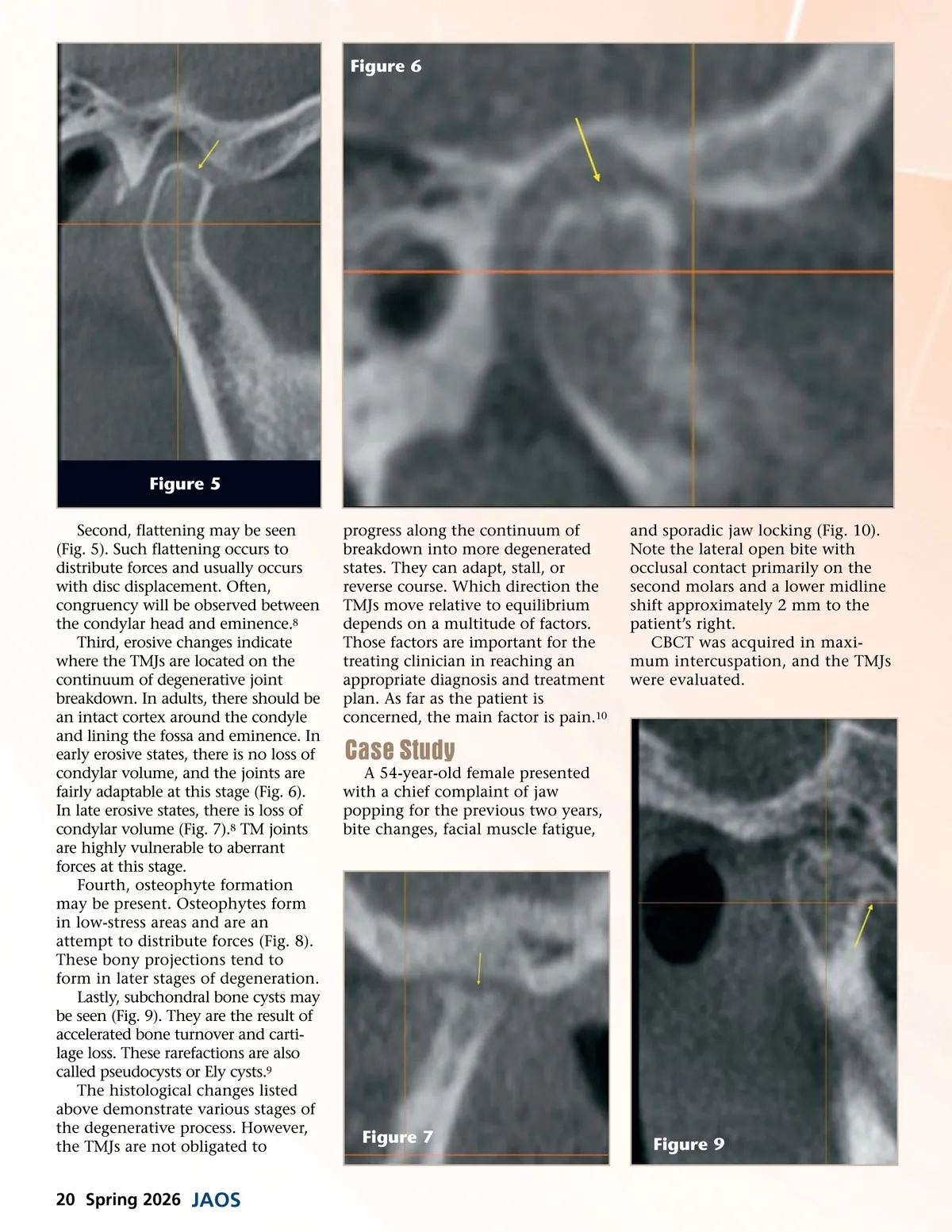
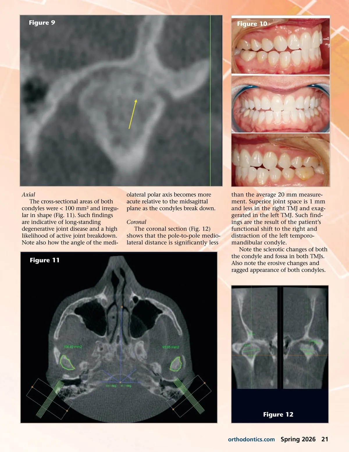
Figure 6 Figure 5 Second, flattening may be seen (Fig. 5). Such flattening occurs to distribute forces and usually occurs with disc displacement. Often, congruency will be observed between the condylar head and eminence. 8 Third, erosive changes indicate where the TMJs are located on the continuum of degenerative joint breakdown. In adults, there should be an intact cortex around the condyle and lining the fossa and eminence. In early erosive states, there is no loss of condylar volume, and the joints are fairly adaptable at this stage (Fig. 6). In late erosive states, there is loss of condylar volume (Fig. 7). 8 TM joints are highly vulnerable to aberrant forces at this stage. Fourth, osteophyte formation may be present. Osteophytes form in low-stress areas and are an attempt to distribute forces (Fig. 8). These bony projections tend to form in later stages of degeneration. Lastly, subchondral bone cysts may be seen (Fig. 9). They are the result of accelerated bone turnover and carti-lage loss. These rarefactions are also called pseudocysts or Ely cysts. 9 The histological changes listed above demonstrate various stages of the degenerative process. However, the TMJs are not obligated to progress along the continuum of breakdown into more degenerated states. They can adapt, stall, or reverse course. Which direction the TMJs move relative to equilibrium depends on a multitude of factors. Those factors are important for the treating clinician in reaching an appropriate diagnosis and treatment plan. As far as the patient is concerned, the main factor is pain. 10 and sporadic jaw locking (Fig. 10). Note the lateral open bite with occlusal contact primarily on the second molars and a lower midline shift approximately 2 mm to the patient’s right. CBCT was acquired in maxi-mum intercuspation, and the TMJs were evaluated. Case Study A 54-year-old female presented with a chief complaint of jaw popping for the previous two years, bite changes, facial muscle fatigue, Figure 7 Figure 9 20 Spring 2026 JAOS
Journal of the American Orthodontic Society Spring 2026: Page 20