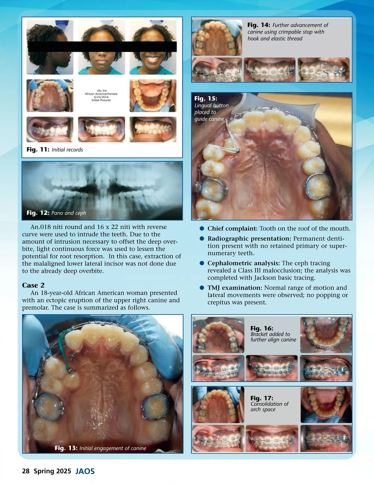
Fig. 19: Final ceph tracing and panographic x-ray Fig. 18: Final composite images b Airway assessment: Normal breathing through the nose was observed, and no history of snoring or obstructive sleep apnea was reported. b Intra-oral examination: Exam revealed an erupted canine between the upper right first and second premolar, Class III molar relationship, and 3 mm upper midline deviation to the right. b Treatment objectives: Level and align all teeth, incorporate ectopic transpositioned tooth into arch alignment, and achieve proper anterior over-bite and overjet, while minimizing arch distortion and root resorption from bodily moving malaligned canine into the proper position. The treatment plan consisted of an SWS MBT prescrip-tion with a .022 slot, full form arch, .012 niti, .014 niti, and .018 niti. Also used were a .020 ss with open coil spring, 16 x 22 niti, 16 x 22 ss, and 18 x 25 ss. Niti wires of .012, .014, and .018 were used for initial leveling and aligning. A .020 ss with an open coil spring was used to widen space for the canine and shift the midline to the left. Once the 18 x 25 wire was placed, attachments were placed on the canine to bring it into the upper arch. The biomechanics to achieve alignment of the canine included everything from elastic threads to power chains; these were used to guide the tooth into alignment until it was close enough to piggyback the 18 x 25 ss with a .014 niti. Fig. 20: Final comparison from occlusal view Case 3 A 40-year-old Cambodian man presented with an ectopic and transposed eruption of the right canine and first premolar. The case is summarized as follows. b Chief complaint: Upper right canine tooth protrusion. b Radiographic presentation: Permanent dentition was present; panographic radiograph revealed the apical portion of the canine was positioned between the premolar teeth. Fig. 21: Initial records www.orthodontics.com Spring 2025 29
Journal of the American Orthodontic Society Spring 2025: Page 29