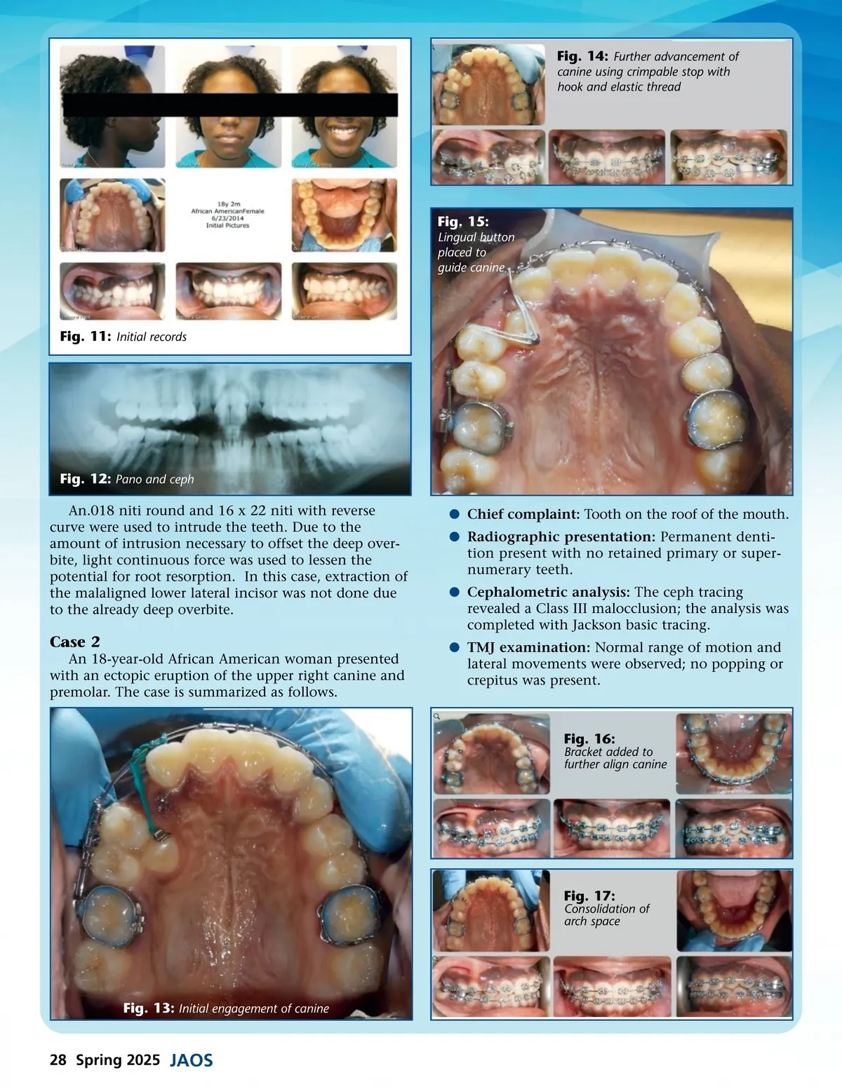
Fig. 14: Further advancement of canine using crimpable stop with hook and elastic thread Fig. 15: Lingual button placed to guide canine Fig. 11: Initial records Fig. 12: Pano and ceph An.018 niti round and 16 x 22 niti with reverse curve were used to intrude the teeth. Due to the amount of intrusion necessary to offset the deep over-bite, light continuous force was used to lessen the potential for root resorption. In this case, extraction of the malaligned lower lateral incisor was not done due to the already deep overbite. b Chief complaint: Tooth on the roof of the mouth. b Radiographic presentation: Permanent denti-tion present with no retained primary or super-numerary teeth. b Cephalometric analysis: The ceph tracing revealed a Class III malocclusion; the analysis was completed with Jackson basic tracing. b TMJ examination: Normal range of motion and lateral movements were observed; no popping or crepitus was present. Case 2 An 18-year-old African American woman presented with an ectopic eruption of the upper right canine and premolar. The case is summarized as follows. Fig. 16: Bracket added to further align canine Fig. 17: Consolidation of arch space Fig. 13: Initial engagement of canine 28 Spring 2025 JAOS
Journal of the American Orthodontic Society Spring 2025: Page 28