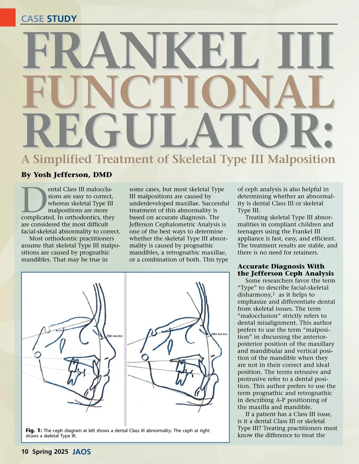
CASE STUDY FUNCTIONAL FRANKEL III some cases, but most skeletal Type III malpositions are caused by underdeveloped maxillae. Successful treatment of this abnormality is based on accurate diagnosis. The Jefferson Cephalometric Analysis is one of the best ways to determine whether the skeletal Type III abnor-mality is caused by prognathic mandibles, a retrognathic maxillae, or a combination of both. This type REGULATOR: A Simplified Treatment of Skeletal Type III Malposition By Yosh Jefferson, DMD ental Class III malocclu-sions are easy to correct, whereas skeletal Type III malpositions are more complicated. In orthodontics, they are considered the most difficult facial-skeletal abnormality to correct. Most orthodontic practitioners assume that skeletal Type III malpo-sitions are caused by prognathic mandibles. That may be true in D of ceph analysis is also helpful in determining whether an abnormal-ity is dental Class III or skeletal Type III. Treating skeletal Type III abnor-malities in compliant children and teenagers using the Frankel III appliance is fast, easy, and efficient. The treatment results are stable, and there is no need for retainers. Accurate Diagnosis With the Jefferson Ceph Analysis Some researchers favor the term “Type” to describe facial-skeletal disharmony, 1 as it helps to emphasize and differentiate dental from skeletal issues. The term “malocclusion” strictly refers to dental misalignment. This author prefers to use the term “malposi-tion” in discussing the anterior-posterior position of the maxillary and mandibular and vertical posi-tion of the mandible when they are not in their correct and ideal position. The terms retrusive and protrusive refer to a dental posi-tion. This author prefers to use the term prognathic and retrognathic in describing A-P positioning of the maxilla and mandible. If a patient has a Class III issue, is it a dental Class III or skeletal Type III? Treating practitioners must know the difference to treat the Fig. 1: The ceph diagram at left shows a dental Class III abnormality. The ceph at right shows a skeletal Type III. 10 Spring 2025 JAOS
Journal of the American Orthodontic Society Spring 2025: Page 10