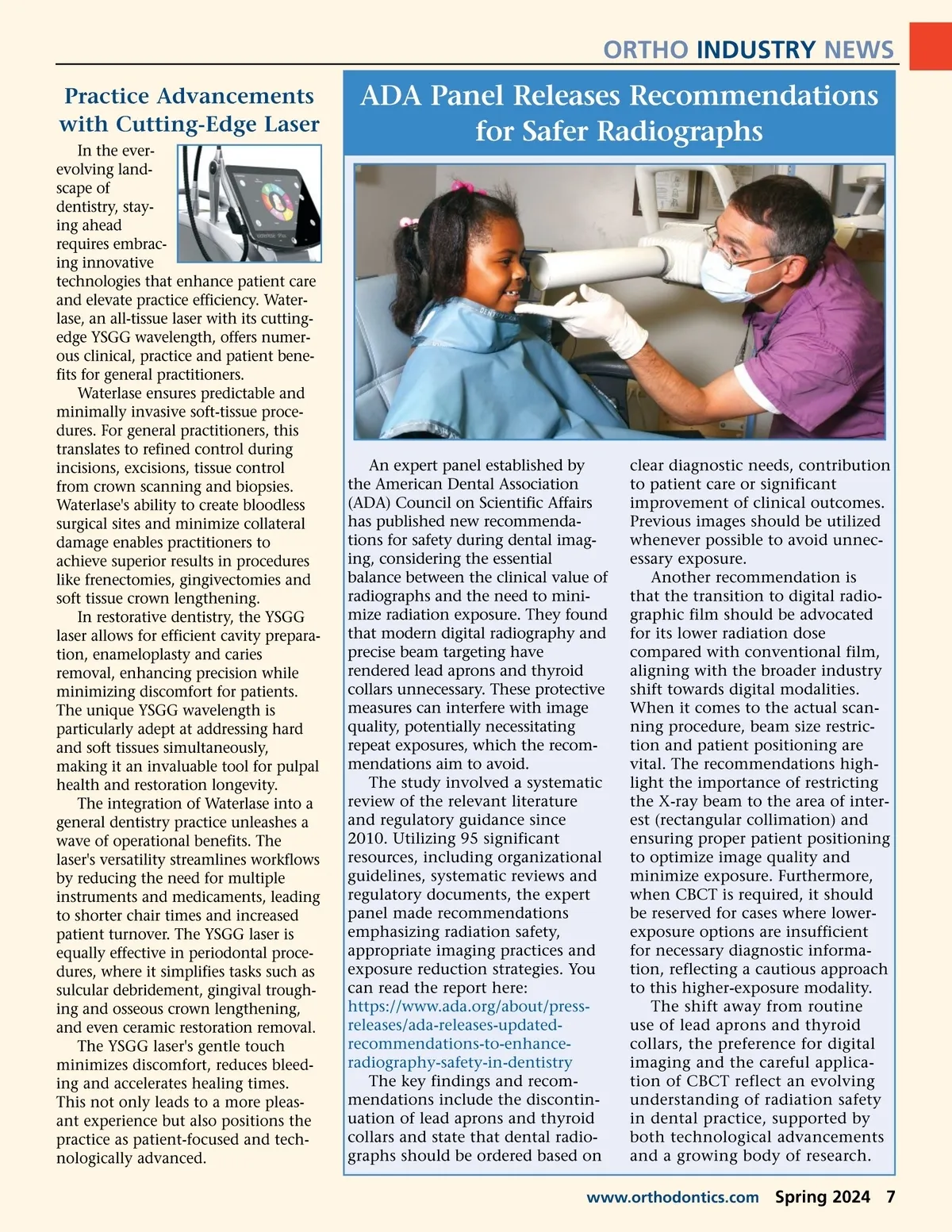
ORTHO INDUSTRY NEWS Practice Advancements with Cutting-Edge Laser In the ever-evolving land-scape of dentistry, stay-ing ahead requires embrac-ing innovative technologies that enhance patient care and elevate practice efficiency. Water-lase, an all-tissue laser with its cutting-edge YSGG wavelength, offers numer-ous clinical, practice and patient bene-fits for general practitioners. Waterlase ensures predictable and minimally invasive soft-tissue proce-dures. For general practitioners, this translates to refined control during incisions, excisions, tissue control from crown scanning and biopsies. Waterlase's ability to create bloodless surgical sites and minimize collateral damage enables practitioners to achieve superior results in procedures like frenectomies, gingivectomies and soft tissue crown lengthening. In restorative dentistry, the YSGG laser allows for efficient cavity prepara-tion, enameloplasty and caries removal, enhancing precision while minimizing discomfort for patients. The unique YSGG wavelength is particularly adept at addressing hard and soft tissues simultaneously, making it an invaluable tool for pulpal health and restoration longevity. The integration of Waterlase into a general dentistry practice unleashes a wave of operational benefits. The laser's versatility streamlines workflows by reducing the need for multiple instruments and medicaments, leading to shorter chair times and increased patient turnover. The YSGG laser is equally effective in periodontal proce-dures, where it simplifies tasks such as sulcular debridement, gingival trough-ing and osseous crown lengthening, and even ceramic restoration removal. The YSGG laser's gentle touch minimizes discomfort, reduces bleed-ing and accelerates healing times. This not only leads to a more pleas-ant experience but also positions the practice as patient-focused and tech-nologically advanced. ADA Panel Releases Recommendations for Safer Radiographs An expert panel established by the American Dental Association (ADA) Council on Scientific Affairs has published new recommenda-tions for safety during dental imag-ing, considering the essential balance between the clinical value of radiographs and the need to mini-mize radiation exposure. They found that modern digital radiography and precise beam targeting have rendered lead aprons and thyroid collars unnecessary. These protective measures can interfere with image quality, potentially necessitating repeat exposures, which the recom-mendations aim to avoid. The study involved a systematic review of the relevant literature and regulatory guidance since 2010. Utilizing 95 significant resources, including organizational guidelines, systematic reviews and regulatory documents, the expert panel made recommendations emphasizing radiation safety, appropriate imaging practices and exposure reduction strategies. You can read the report here: https://www.ada.org/about/press-releases/ada-releases-updated-recommendations-to-enhance-radiography-safety-in-dentistry The key findings and recom-mendations include the discontin-uation of lead aprons and thyroid collars and state that dental radio-graphs should be ordered based on clear diagnostic needs, contribution to patient care or significant improvement of clinical outcomes. Previous images should be utilized whenever possible to avoid unnec-essary exposure. Another recommendation is that the transition to digital radio-graphic film should be advocated for its lower radiation dose compared with conventional film, aligning with the broader industry shift towards digital modalities. When it comes to the actual scan-ning procedure, beam size restric-tion and patient positioning are vital. The recommendations high-light the importance of restricting the X-ray beam to the area of inter-est (rectangular collimation) and ensuring proper patient positioning to optimize image quality and minimize exposure. Furthermore, when CBCT is required, it should be reserved for cases where lower-exposure options are insufficient for necessary diagnostic informa-tion, reflecting a cautious approach to this higher-exposure modality. The shift away from routine use of lead aprons and thyroid collars, the preference for digital imaging and the careful applica-tion of CBCT reflect an evolving understanding of radiation safety in dental practice, supported by both technological advancements and a growing body of research. www.orthodontics.com Spring 2024 7
Journal of the American Orthodontic Society Spring 2024: Page 7