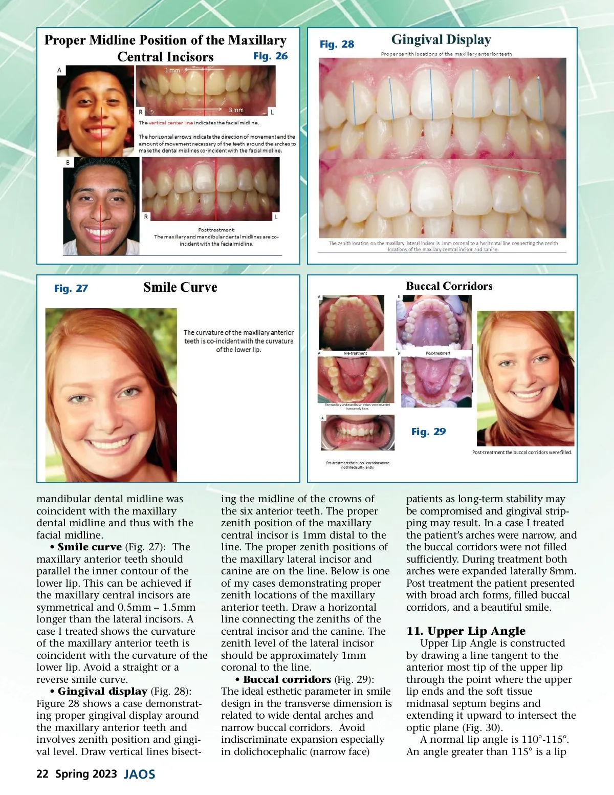
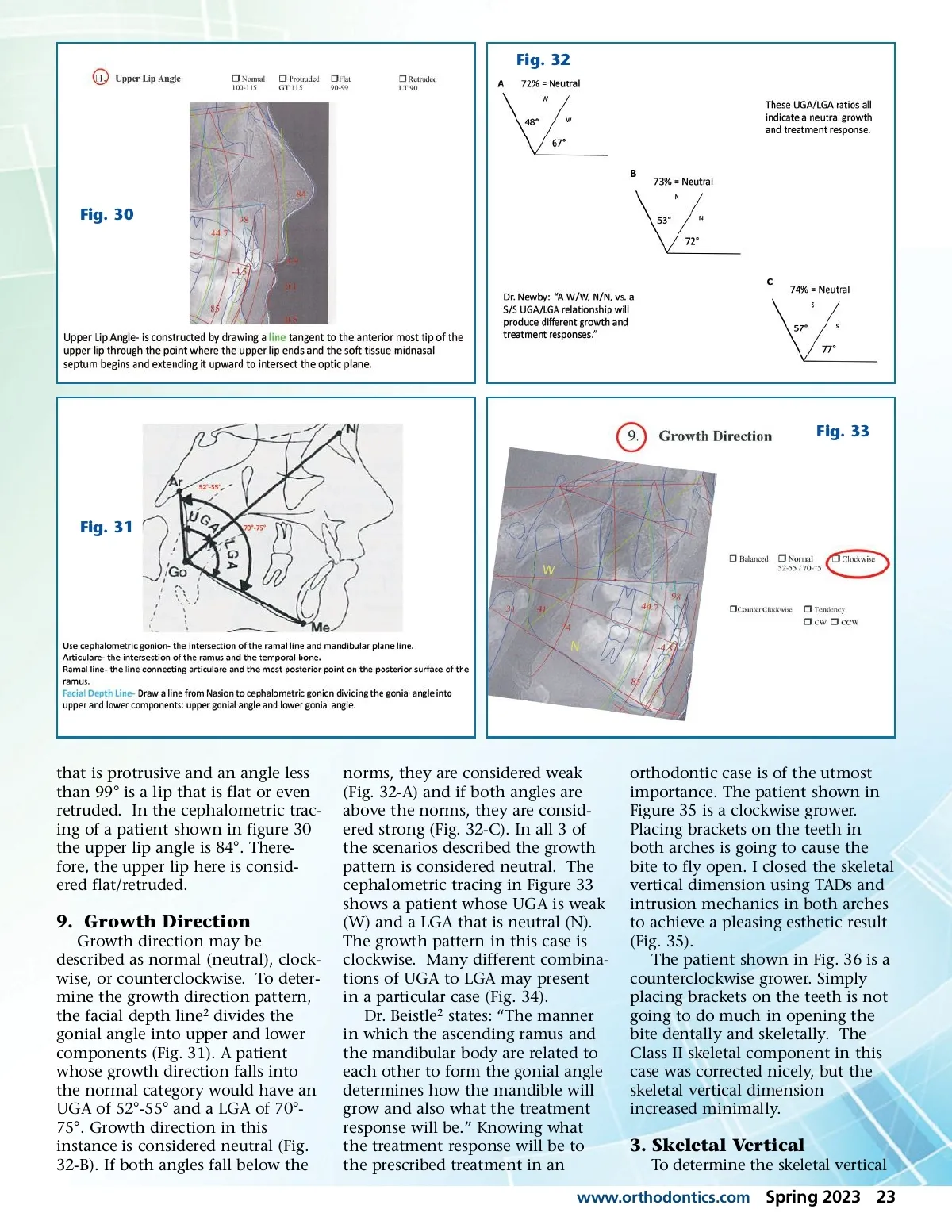
Fig. 32 Fig. 30 Fig. 33 Fig. 31 that is protrusive and an angle less than 99° is a lip that is flat or even retruded. In the cephalometric trac-ing of a patient shown in figure 30 the upper lip angle is 84°. There-fore, the upper lip here is consid-ered flat/retruded. 9. Growth Direction Growth direction may be described as normal (neutral), clock-wise, or counterclockwise. To deter-mine the growth direction pattern, the facial depth line 2 divides the gonial angle into upper and lower components (Fig. 31). A patient whose growth direction falls into the normal category would have an UGA of 52°-55° and a LGA of 70°-75°. Growth direction in this instance is considered neutral (Fig. 32-B). If both angles fall below the norms, they are considered weak (Fig. 32-A) and if both angles are above the norms, they are consid-ered strong (Fig. 32-C). In all 3 of the scenarios described the growth pattern is considered neutral. The cephalometric tracing in Figure 33 shows a patient whose UGA is weak (W) and a LGA that is neutral (N). The growth pattern in this case is clockwise. Many different combina-tions of UGA to LGA may present in a particular case (Fig. 34). Dr. Beistle 2 states: “The manner in which the ascending ramus and the mandibular body are related to each other to form the gonial angle determines how the mandible will grow and also what the treatment response will be.” Knowing what the treatment response will be to the prescribed treatment in an orthodontic case is of the utmost importance. The patient shown in Figure 35 is a clockwise grower. Placing brackets on the teeth in both arches is going to cause the bite to fly open. I closed the skeletal vertical dimension using TADs and intrusion mechanics in both arches to achieve a pleasing esthetic result (Fig. 35). The patient shown in Fig. 36 is a counterclockwise grower. Simply placing brackets on the teeth is not going to do much in opening the bite dentally and skeletally. The Class II skeletal component in this case was corrected nicely, but the skeletal vertical dimension increased minimally. 3. Skeletal Vertical To determine the skeletal vertical www.orthodontics.com Spring 2023 23
Journal of the American Orthodontic Society Spring 2023: Page 23