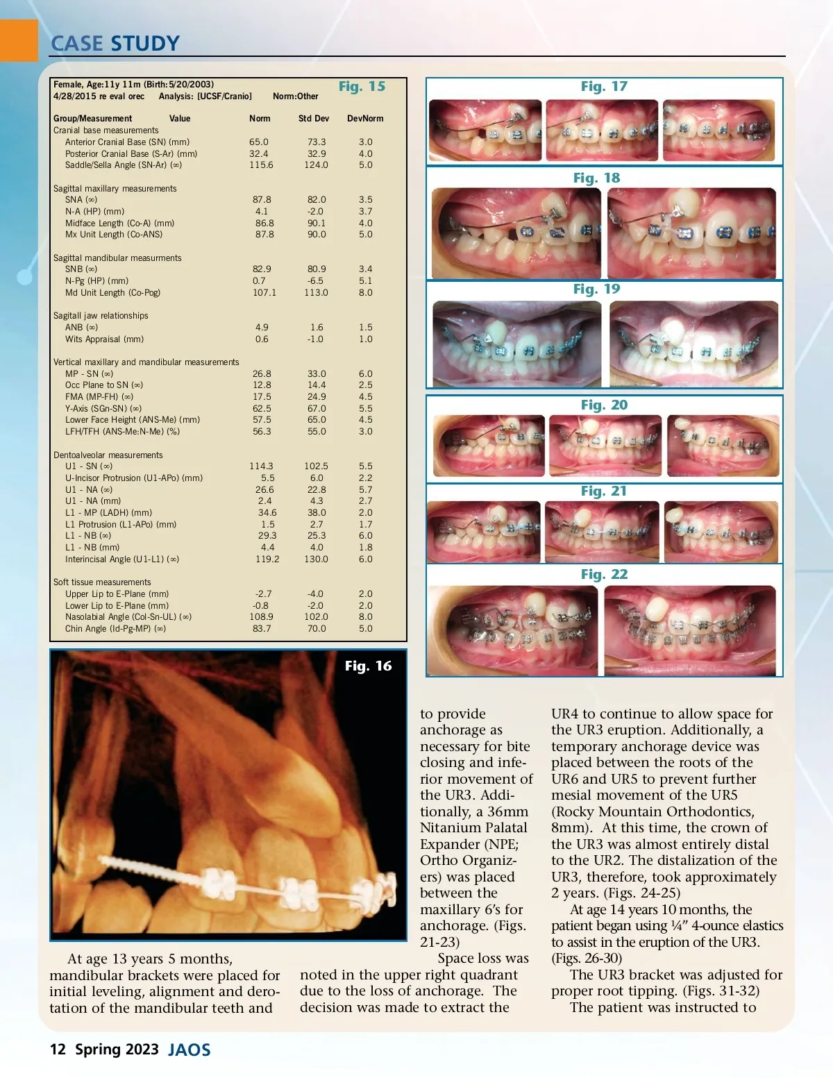
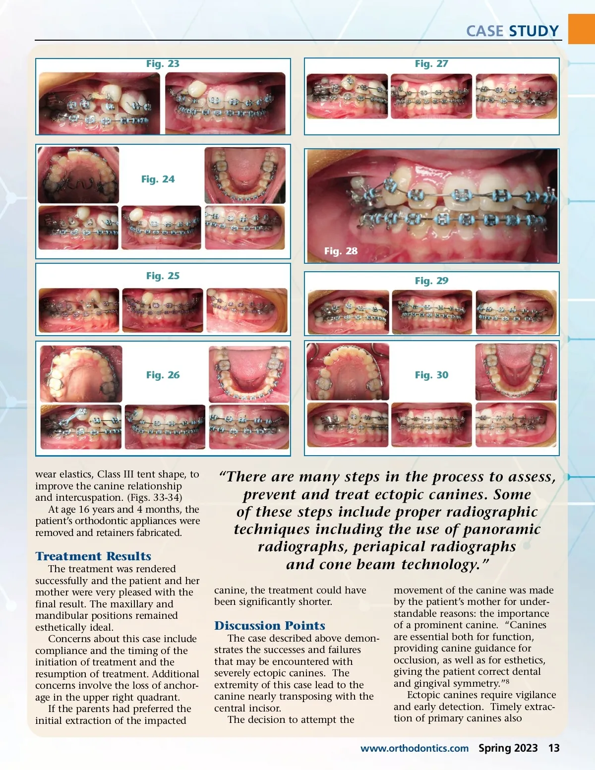
CASE STUDY Female, Age:11y 11m (Birth:5/20/2003) 4/28/2015 re eval orec Analysis: [UCSF/Cranio] Group/Measurement Value Cranial base measurements Anterior Cranial Base (SN) (mm) Posterior Cranial Base (S-Ar) (mm) Saddle/Sella Angle (SN-Ar) ( ∞ ) Sagittal maxillary measurements SNA ( ∞ ) N-A (HP) (mm) Midface Length (Co-A) (mm) Mx Unit Length (Co-ANS) Sagittal mandibular measurments SNB ( ∞ ) N-Pg (HP) (mm) Md Unit Length (Co-Pog) Sagitall jaw relationships ANB ( ∞ ) Wits Appraisal (mm) Vertical maxillary and mandibular measurements MP -SN ( ∞ ) Occ Plane to SN ( ∞ ) FMA (MP-FH) ( ∞ ) Y-Axis (SGn-SN) ( ∞ ) Lower Face Height (ANS-Me) (mm) LFH/TFH (ANS-Me:N-Me) (%) Dentoalveolar measurements U1 -SN ( ∞ ) U-Incisor Protrusion (U1-APo) (mm) U1 -NA ( ∞ ) U1 -NA (mm) L1 -MP (LADH) (mm) L1 Protrusion (L1-APo) (mm) L1 -NB ( ∞ ) L1 -NB (mm) Interincisal Angle (U1-L1) ( ∞ ) Soft tissue measurements Upper Lip to E-Plane (mm) Lower Lip to E-Plane (mm) Nasolabial Angle (Col-Sn-UL) ( ∞ ) Chin Angle (Id-Pg-MP) ( ∞ ) Norm 65.0 32.4 115.6 Norm:Other Std Dev 73.3 32.9 124.0 Fig. 15 DevNorm 3.0 4.0 5.0 Fig. 17 Fig. 18 87.8 4.1 86.8 87.8 82.0 -2.0 90.1 90.0 3.5 3.7 4.0 5.0 82.9 0.7 107.1 80.9 -6.5 113.0 3.4 5.1 8.0 Fig. 19 4.9 0.6 1.6 -1.0 1.5 1.0 26.8 12.8 17.5 62.5 57.5 56.3 33.0 14.4 24.9 67.0 65.0 55.0 6.0 2.5 4.5 5.5 4.5 3.0 Fig. 20 114.3 5.5 26.6 2.4 34.6 1.5 29.3 4.4 119.2 102.5 6.0 22.8 4.3 38.0 2.7 25.3 4.0 130.0 5.5 2.2 5.7 2.7 2.0 1.7 6.0 1.8 6.0 Fig. 21 Fig. 22 -2.7 -0.8 108.9 83.7 -4.0 -2.0 102.0 70.0 2.0 2.0 8.0 5.0 Fig. 16 At age 13 years 5 months, mandibular brackets were placed for initial leveling, alignment and dero-tation of the mandibular teeth and to provide anchorage as necessary for bite closing and infe-rior movement of the UR3. Addi-tionally, a 36mm Nitanium Palatal Expander (NPE; Ortho Organiz-ers) was placed between the maxillary 6’s for anchorage. (Figs. 21-23) Space loss was noted in the upper right quadrant due to the loss of anchorage. The decision was made to extract the UR4 to continue to allow space for the UR3 eruption. Additionally, a temporary anchorage device was placed between the roots of the UR6 and UR5 to prevent further mesial movement of the UR5 (Rocky Mountain Orthodontics, 8mm). At this time, the crown of the UR3 was almost entirely distal to the UR2. The distalization of the UR3, therefore, took approximately 2 years. (Figs. 24-25) At age 14 years 10 months, the patient began using ¼” 4-ounce elastics to assist in the eruption of the UR3. (Figs. 26-30) The UR3 bracket was adjusted for proper root tipping. (Figs. 31-32) The patient was instructed to 12 Spring 2023 JAOS
Journal of the American Orthodontic Society Spring 2023: Page 12