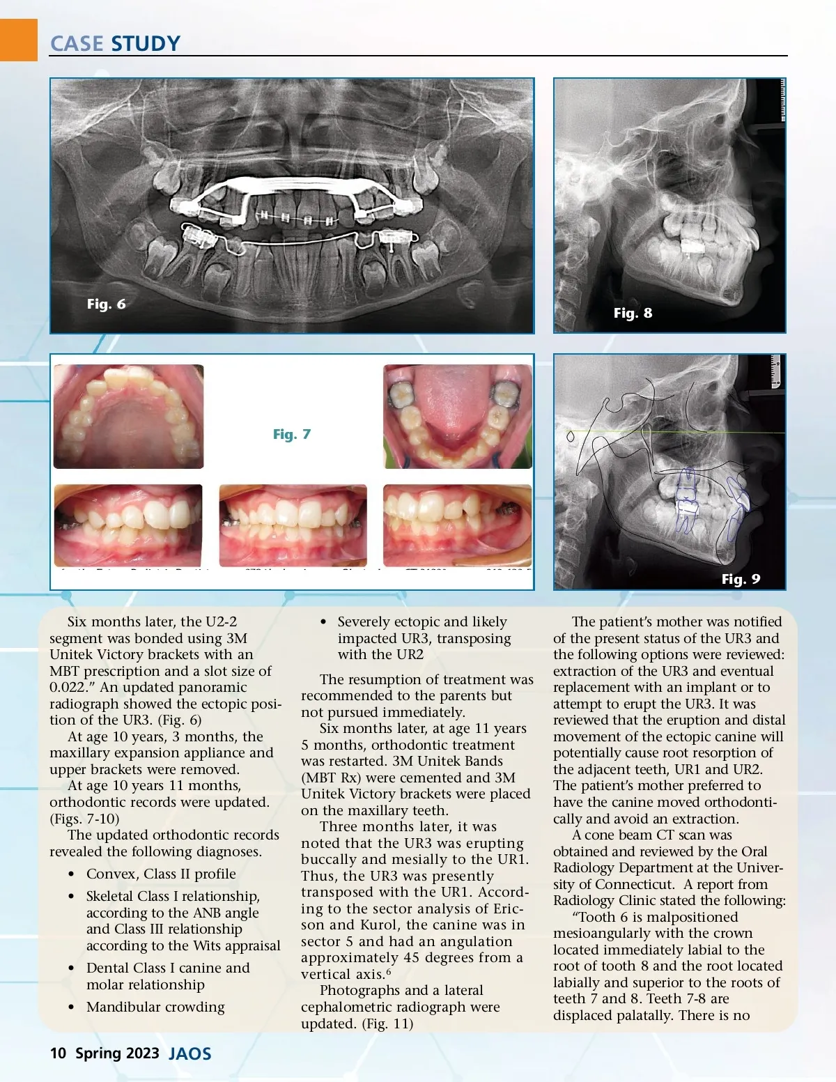
CASE STUDY Female, Age:10y 11m (Birth:5/20/2003) 4/29/2014 Phase II Analysis: [UCSF/Cranio] Group/Measurement Value Cranial base measurements Anterior Cranial Base (SN) (mm) Posterior Cranial Base (S-Ar) (mm) Saddle/Sella Angle (SN-Ar) ( ∞ ) Sagittal maxillary measurements SNA ( ∞ ) N-A (HP) (mm) Midface Length (Co-A) (mm) Mx Unit Length (Co-ANS) Sagittal mandibular measurments SNB ( ∞ ) N-Pg (HP) (mm) Md Unit Length (Co-Pog) Sagitall jaw relationships ANB ( ∞ ) Wits Appraisal (mm) Vertical maxillary and mandibular measurements MP -SN ( ∞ ) Occ Plane to SN ( ∞ ) FMA (MP-FH) ( ∞ ) Y-Axis (SGn-SN) ( ∞ ) Lower Face Height (ANS-Me) (mm) LFH/TFH (ANS-Me:N-Me) (%) Dentoalveolar measurements U1 -SN ( ∞ ) U-Incisor Protrusion (U1-APo) (mm) U1 -NA ( ∞ ) U1 -NA (mm) L1 -MP (LADH) (mm) L1 Protrusion (L1-APo) (mm) L1 -NB ( ∞ ) L1 -NB (mm) Interincisal Angle (U1-L1) ( ∞ ) Soft tissue measurements Upper Lip to E-Plane (mm) Lower Lip to E-Plane (mm) Nasolabial Angle (Col-Sn-UL) ( ∞ ) Chin Angle (Id-Pg-MP) ( ∞ ) Norm:Other Norm 64.4 27.9 122.5 Std Dev 72.6 32.3 124.0 Fig. 10 DevNorm 3.0 4.0 5.0 82.3 -0.6 76.1 81.1 82.0 -2.0 89.1 90.0 3.5 3.7 4.0 5.0 81.2 4.0 96.6 80.9 -6.5 113.0 3.4 5.1 8.0 1.0 -3.8 1.6 -1.0 1.5 1.0 30.9 17.6 20.5 63.6 54.4 54.7 33.0 14.4 25.2 67.0 65.0 55.0 6.0 2.5 4.5 5.5 4.5 3.0 Fig. 12 Fig. 13 113.2 7.8 30.9 6.7 33.0 3.6 22.3 3.8 125.8 102.4 6.0 22.8 4.3 38.0 2.7 25.3 4.0 130.0 5.5 2.2 5.7 2.7 2.0 1.7 6.0 1.8 6.0 -0.6 1.0 113.2 79.9 -3.3 -2.0 102.0 70.0 2.0 2.0 8.0 5.0 Fig. 14 Fig. 11 resorption of the surrounding teeth.” (Figs. 13-15) We reviewed that treatment of the partially transposed and impacted canine would be halted if root resorption was noted on adjoining teeth. At age 12 years 4 months, move-ment of the impacted canine was begun using a cantilever/whip spring. A 0.018x0.025” stainless steel wire was bent with a mesial stop adjacent to the UR6 tube and a helix placed at the mesial end for ligation. An eruption loop was bonded to the UR1. A buccal and inferior force was placed on the UR1 for the purpose of moving the crown away from the adjoining teeth. That action was continued for 5 months. An archwire was purposefully not engaged in the maxillary anterior teeth in order to allow the teeth to move at will during the eruptive process of the UR3. At times, only a steel ligature wire was used to prevent distal drift-ing. (Fig. 16) The existing cantilever spring continued to be activated inferiorly and buccally and tied to the bonded eruption loop with elastic thread (Dynaflex). (Figs. 17-20) www.orthodontics.com Spring 2023 11
Journal of the American Orthodontic Society Spring 2023: Page 11