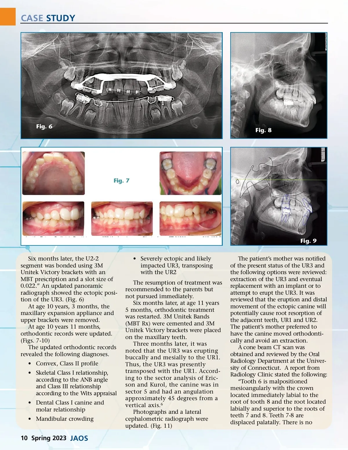
CASE STUDY Fig. 6 Fig. 8 Fig. 7 Fig. 9 Six months later, the U2-2 segment was bonded using 3M Unitek Victory brackets with an MBT prescription and a slot size of 0.022.” An updated panoramic radiograph showed the ectopic posi-tion of the UR3. (Fig. 6) At age 10 years, 3 months, the maxillary expansion appliance and upper brackets were removed. At age 10 years 11 months, orthodontic records were updated. (Figs. 7-10) The updated orthodontic records revealed the following diagnoses. • Convex, Class II profile • Skeletal Class I relationship, according to the ANB angle and Class III relationship according to the Wits appraisal • Dental Class I canine and molar relationship • Mandibular crowding • Severely ectopic and likely impacted UR3, transposing with the UR2 The resumption of treatment was recommended to the parents but not pursued immediately. Six months later, at age 11 years 5 months, orthodontic treatment was restarted. 3M Unitek Bands (MBT Rx) were cemented and 3M Unitek Victory brackets were placed on the maxillary teeth. Three months later, it was noted that the UR3 was erupting buccally and mesially to the UR1. Thus, the UR3 was presently transposed with the UR1. Accord-ing to the sector analysis of Eric-son and Kurol, the canine was in sector 5 and had an angulation approximately 45 degrees from a vertical axis. 6 Photographs and a lateral cephalometric radiograph were updated. (Fig. 11) The patient’s mother was notified of the present status of the UR3 and the following options were reviewed: extraction of the UR3 and eventual replacement with an implant or to attempt to erupt the UR3. It was reviewed that the eruption and distal movement of the ectopic canine will potentially cause root resorption of the adjacent teeth, UR1 and UR2. The patient’s mother preferred to have the canine moved orthodonti-cally and avoid an extraction. A cone beam CT scan was obtained and reviewed by the Oral Radiology Department at the Univer-sity of Connecticut. A report from Radiology Clinic stated the following: “Tooth 6 is malpositioned mesioangularly with the crown located immediately labial to the root of tooth 8 and the root located labially and superior to the roots of teeth 7 and 8. Teeth 7-8 are displaced palatally. There is no 10 Spring 2023 JAOS
Journal of the American Orthodontic Society Spring 2023: Page 10