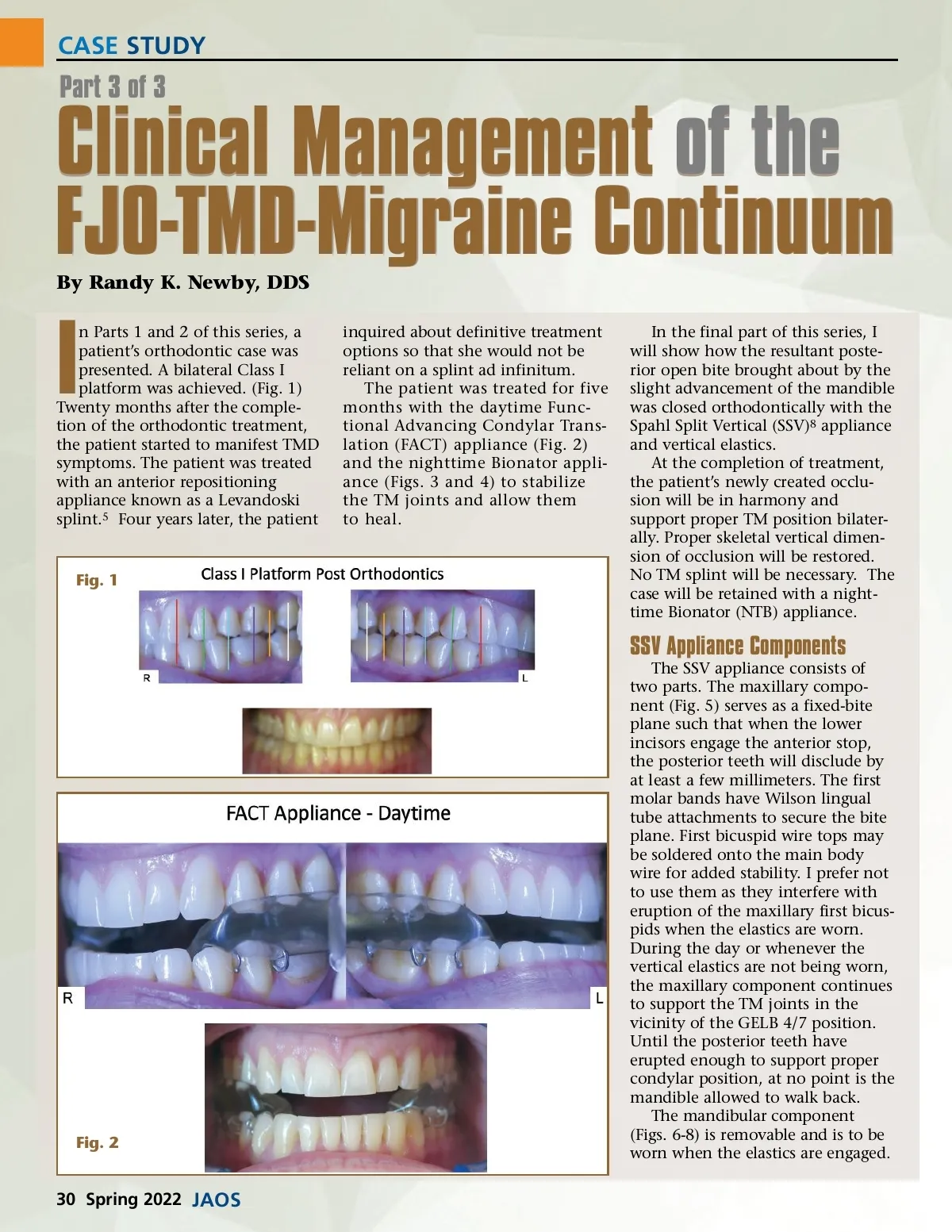
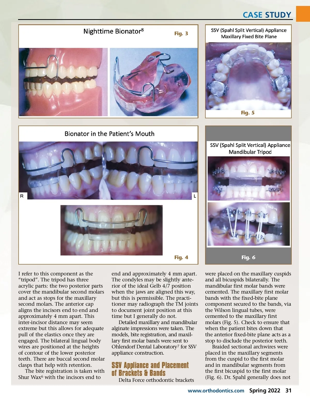
CASE STUDY Fig. 3 Fig. 5 Fig. 4 I refer to this component as the “tripod”. The tripod has three acrylic parts: the two posterior parts cover the mandibular second molars and act as stops for the maxillary second molars. The anterior cap aligns the incisors end to end and approximately 4 mm apart. This inter-incisor distance may seem extreme but this allows for adequate pull of the elastics once they are engaged. The bilateral lingual body wires are positioned at the heights of contour of the lower posterior teeth. There are buccal second molar clasps that help with retention. The bite registration is taken with Shur Wax 6 with the incisors end to end and approximately 4 mm apart. The condyles may be slightly ante-rior of the ideal Gelb 4/7 position when the jaws are aligned this way, but this is permissible. The practi-tioner may radiograph the TM joints to document joint position at this time but I generally do not. Detailed maxillary and mandibular alginate impressions were taken. The models, bite registration, and maxil-lary first molar bands were sent to Ohlendorf Dental Laboratory 7 for SSV appliance construction. Fig. 6 were placed on the maxillary cuspids and all bicuspids bilaterally. The mandibular first molar bands were cemented. The maxillary first molar bands with the fixed-bite plane component secured to the bands, via the Wilson lingual tubes, were cemented to the maxillary first molars (Fig. 5). Check to ensure that when the patient bites down that the anterior fixed-bite plane acts as a stop to disclude the posterior teeth. Braided sectional archwires were placed in the maxillary segments from the cuspid to the first molar and in mandibular segments from the first bicuspid to the first molar (Fig. 6). Dr. Spahl generally does not SSV Appliance and Placement of Brackets & Bands Delta Force orthodontic brackets www.orthodontics.com Spring 2022 31
Journal of the American Orthodontic Society Spring 2022: Page 31