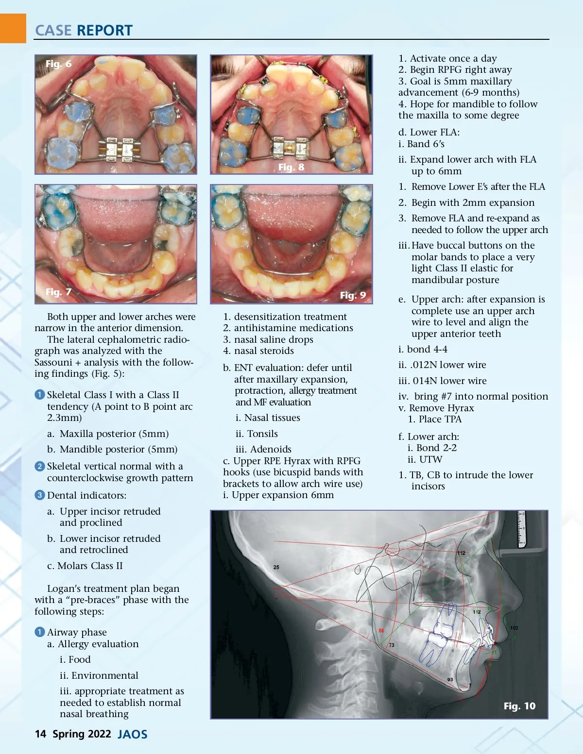
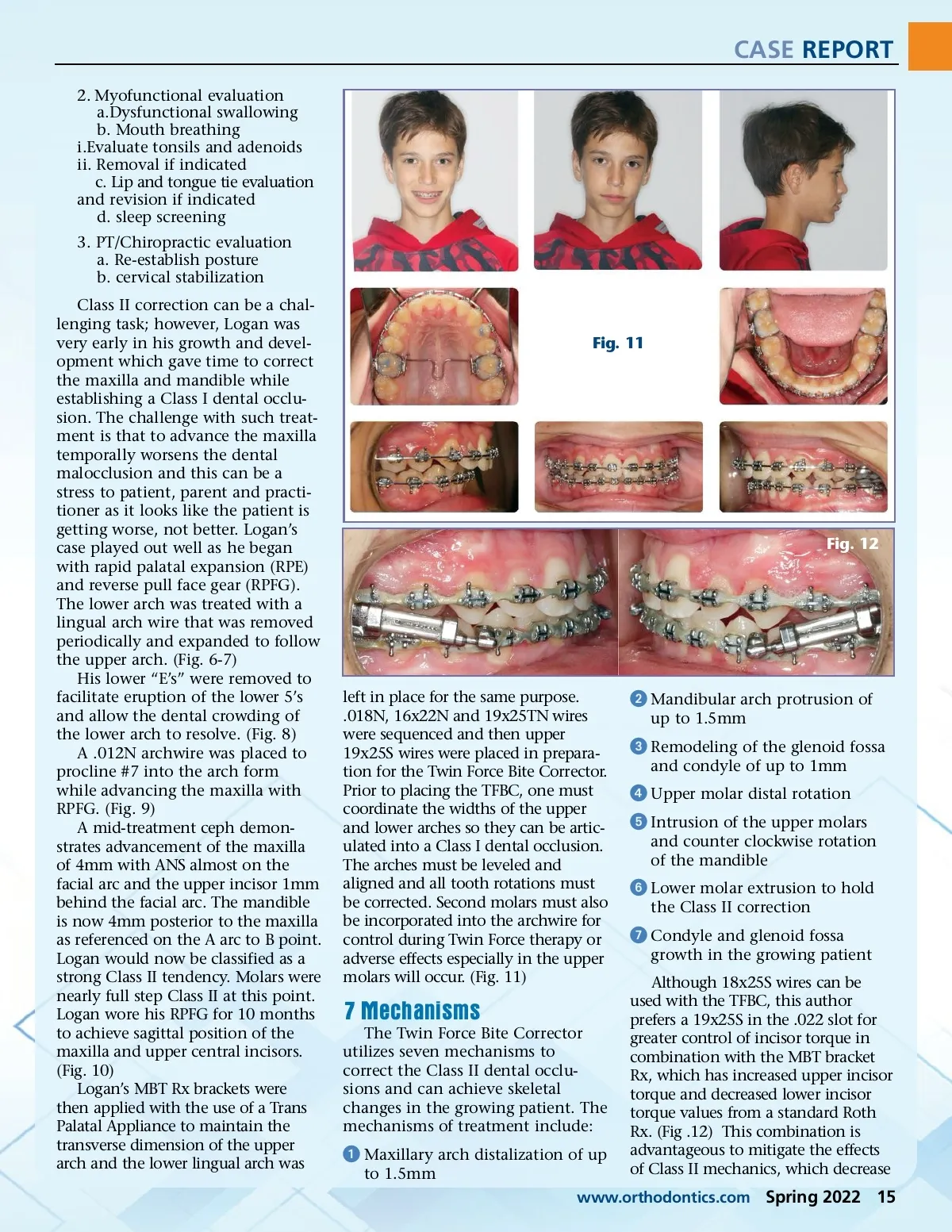
CASE REPORT Fig. 6 1. Activate once a day 2. Begin RPFG right away 3. Goal is 5mm maxillary advancement (6-9 months) 4. Hope for mandible to follow the maxilla to some degree d. Lower FLA: i. Band 6’s Fig. 8 ii. Expand lower arch with FLA up to 6mm 1. Remove Lower E’s after the FLA 2. Begin with 2mm expansion 3. Remove FLA and re-expand as needed to follow the upper arch iii. Have buccal buttons on the molar bands to place a very light Class II elastic for mandibular posture Fig. 7 Both upper and lower arches were narrow in the anterior dimension. The lateral cephalometric radio-graph was analyzed with the Sassouni + analysis with the follow-ing findings (Fig. 5): 1. 2. 3. 4. Fig. 9 desensitization treatment antihistamine medications nasal saline drops nasal steroids e. Upper arch: after expansion is complete use an upper arch wire to level and align the upper anterior teeth i. bond 4-4 ii. .012N lower wire iii. 014N lower wire iv. bring #7 into normal position v. Remove Hyrax 1. Place TPA f. Lower arch: i. Bond 2-2 ii. UTW 1. TB, CB to intrude the lower incisors ᕡ Skeletal Class I with a Class II tendency (A point to B point arc 2.3mm) a. Maxilla posterior (5mm) b. Mandible posterior (5mm) b. ENT evaluation: defer until after maxillary expansion, protraction, allergy treatment and MF evaluation i. Nasal tissues ii. Tonsils iii. Adenoids c. Upper RPE Hyrax with RPFG hooks (use bicuspid bands with brackets to allow arch wire use) i. Upper expansion 6mm ᕢ Skeletal vertical normal with a counterclockwise growth pattern ᕣ Dental indicators: a. Upper incisor retruded and proclined b. Lower incisor retruded and retroclined c. Molars Class II Logan’s treatment plan began with a “pre-braces” phase with the following steps: ᕡ Airway phase a. Allergy evaluation i. Food ii. Environmental iii. appropriate treatment as needed to establish normal nasal breathing Fig. 10 14 Spring 2022 JAOS
Journal of the American Orthodontic Society Spring 2022: Page 14