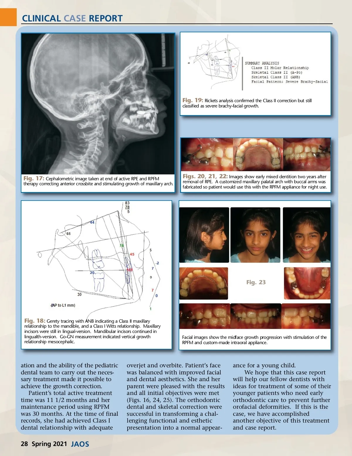
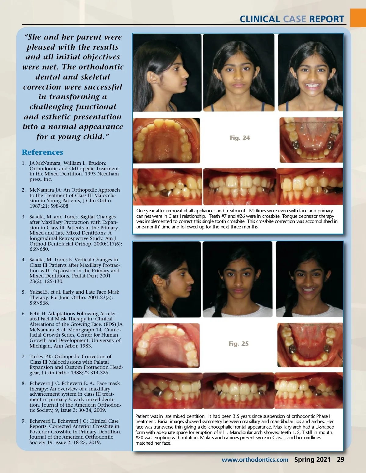
CLINICAL CASE REPORT “She and her parent were pleased with the results and all initial objectives were met. The orthodontic dental and skeletal correction were successful in transforming a challenging functional and esthetic presentation into a normal appearance for a young child.” References 1. JA McNamara, William L. Brudon: Orthodontic and Orthopedic Treatment in the Mixed Dentition. 1993 Needham press, Inc. 2. McNamara JA: An Orthopedic Approach to the Treatment of Class III Malocclu-sion in Young Patients, J Clin Ortho 1987;21: 598-608 3. Saadia, M. and Torres, Sagital Changes after Maxillary Protraction with Expan-sion in Class III Patients in the Primary, Mixed and Late Mixed Dentitions: A longitudinal Retrospective Study. Am J Orthod Dentofacial Orthop. 2000:117(6): 669-680. 4. Saadia, M. Torres,E. Vertical Changes in Class III Patients after Maxillary Protrac-tion with Expansion in the Primary and Mixed Dentitions. Pediat Dent 2001 23(2): 125-130. 5. Yuksel.S. et al. Early and Late Face Mask Therapy. Eur Jour. Ortho. 2001;23(5): 539-568. 6. Petit H: Adaptations Following Acceler-ated Facial Mask Therapy in: Clinical Alterations of the Growing Face. (EDS) JA McNamara et al. Monograph 14, Cranio-facial Growth Series, Center for Human Growth and Development, University of Michigan, Ann Arbor, 1983. 7. Turley P.K: Orthopedic Correction of Class III Malocclusions with Palatal Expansion and Custom Protraction Head-gear, J Clin Ortho 1988;22 314-325. 8. Echeverri J C, Echeverri E. A.: Face mask therapy: An overview of a maxillary advancement system in class III treat-ment in primary & early mixed denti-tion. Journal of the American Orthodon-tic Society, 9, issue 3: 30-34, 2009. 9. Echeverri E, Echeverri J C: Clinical Case Reports: Corrected Anterior Crossbite in Posterior Crossbite in Primary Dentition. Journal of the American Orthodontic Society 19, issue 2: 18-25, 2019. Patient was in late mixed dentition. It had been 3.5 years since suspension of orthodontic Phase I treatment. Facial images showed symmetry between maxillary and mandibular lips and arches. Her face was transverse thin giving a dolichocephalic frontal appearance. Maxillary arch had a U-shaped form with adequate space for eruption of #11. Mandibular arch showed teeth L, S, T still in mouth. #20 was erupting with rotation. Molars and canines present were in Class I, and her midlines matched her face. Fig. 24 One year after removal of all appliances and treatment. Midlines were even with face and primary canines were in Class I relationship. Teeth #7 and #26 were in crossbite. Tongue depressor therapy was implemented to correct this single tooth crossbite. This crossbite correction was accomplished in one-month’ time and followed up for the next three months. Fig. 25 www.orthodontics.com Spring 2021 29
Journal of the American Orthodontic Society Spring 2021: Page 29