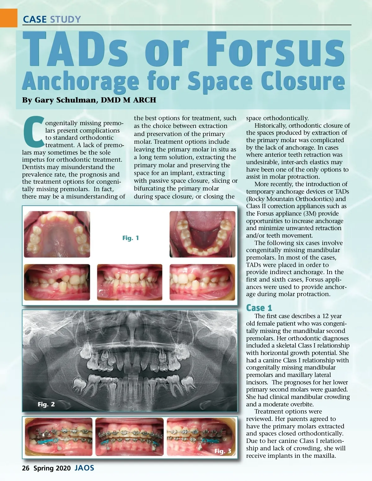
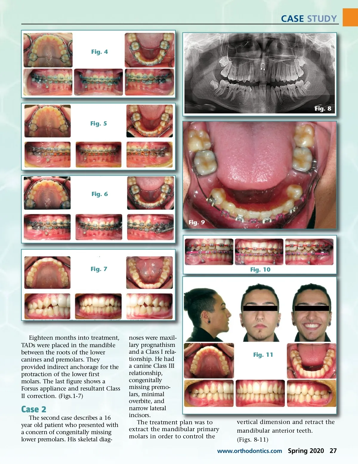
CASE STUDY Fig. 4 Fig. 8 Fig. 5 Fig. 6 Fig. 9 Fig. 7 Fig. 10 Eighteen months into treatment, TADs were placed in the mandible between the roots of the lower canines and premolars. They provided indirect anchorage for the protraction of the lower first molars. The last figure shows a Forsus appliance and resultant Class II correction. (Figs.1-7) Case 2 The second case describes a 16 year old patient who presented with a concern of congenitally missing lower premolars. His skeletal diag-noses were maxil-lary prognathism and a Class I rela-tionship. He had a canine Class III relationship, congenitally missing premo-lars, minimal overbite, and narrow lateral incisors. The treatment plan was to extract the mandibular primary molars in order to control the Fig. 11 vertical dimension and retract the mandibular anterior teeth. (Figs. 8-11) www.orthodontics.com Spring 2020 27
Journal of the American Orthodontic Society Spring 2020: Page 27