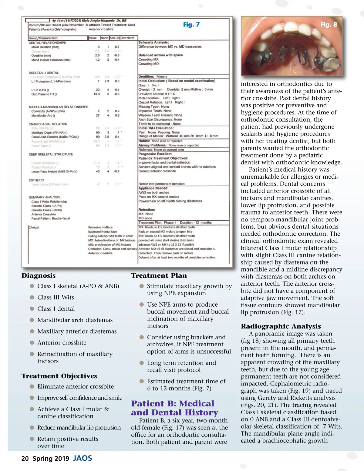
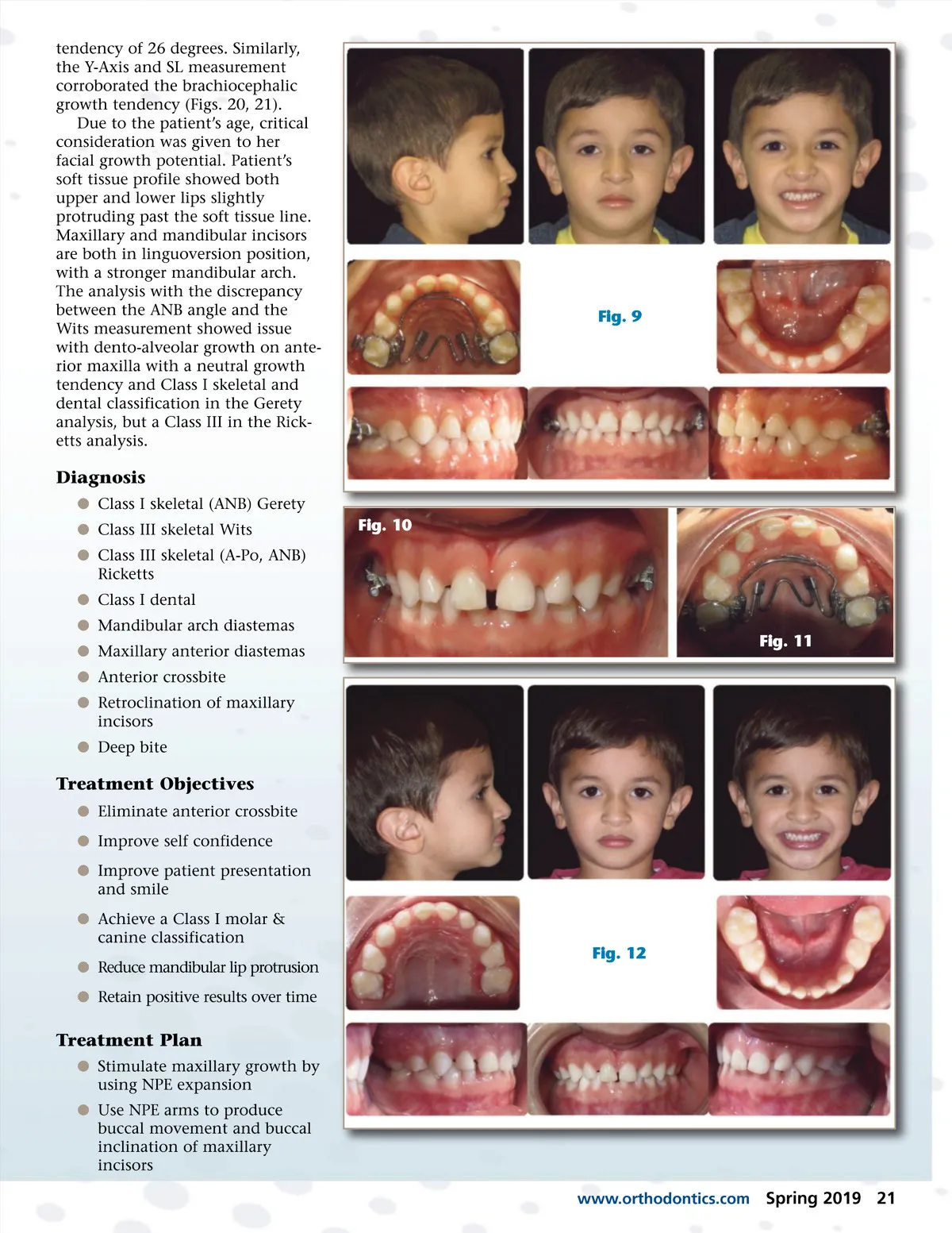
Fig. 7 Fig. 8 Diagnosis b Class I skeletal (A-PO & ANB) b Class III Wits b Class I dental b Mandibular arch diastemas b Maxillary anterior diastemas b Anterior crossbite b Retroclination of maxillary incisors Treatment Plan b Stimulate maxillary growth by using NPE expansion b Use NPE arms to produce buccal movement and buccal inclination of maxillary incisors b Consider using brackets and archwires, if NPE treatment option of arms is unsuccessful b Long term retention and recall visit protocol b Estimated treatment time of 6 to 12 months (Fig. 7) interested in orthodontics due to their awareness of the patient’s ante-rior crossbite. Past dental history was positive for preventive and hygiene procedures. At the time of orthodontic consultation, the patient had previously undergone sealants and hygiene procedures with her treating dentist, but both parents wanted the orthodontic treatment done by a pediatric dentist with orthodontic knowledge. Patient’s medical history was unremarkable for allergies or medi-cal problems. Dental concerns included anterior crossbite of all incisors and mandibular canines, lower lip protrusion, and possible trauma to anterior teeth. There were no temporo-mandibular joint prob-lems, but obvious dental situations needed orthodontic correction. The clinical orthodontic exam revealed bilateral Class I molar relationship with slight Class III canine relation-ship caused by diastema on the mandible and a midline discrepancy with diastemas on both arches on anterior teeth. The anterior cross-bite did not have a component of adaptive jaw movement. The soft tissue contours showed mandibular lip protrusion (Fig. 17). Radiographic Analysis A panoramic image was taken (fig 18) showing all primary teeth present in the mouth, and perma-nent teeth forming. There is an apparent crowding of the maxillary teeth, but due to the young age permanent teeth are not considered impacted. Cephalometric radio-graph was taken (Fig. 19) and traced using Gerety and Ricketts analysis (Figs. 20, 21). The tracing revealed Class I skeletal classification based on 0 ANB and a Class III dentoalve-olar skeletal classification of -7 Wits. The mandibular plane angle indi-cated a brachiocephalic growth Treatment Objectives b Eliminate anterior crossbite b Improve self confidence and smile b Achieve a Class I molar & canine classification b Reduce mandibular lip protrusion b Retain positive results over time Patient B: Medical and Dental History Patient B, a six-year, two-month-old female (Fig. 17) was seen at the office for an orthodontic consulta-tion. Both patient and parent were 20 Spring 2019 JAOS
Journal of the American Orthodontic Society Spring 2019: Page 20