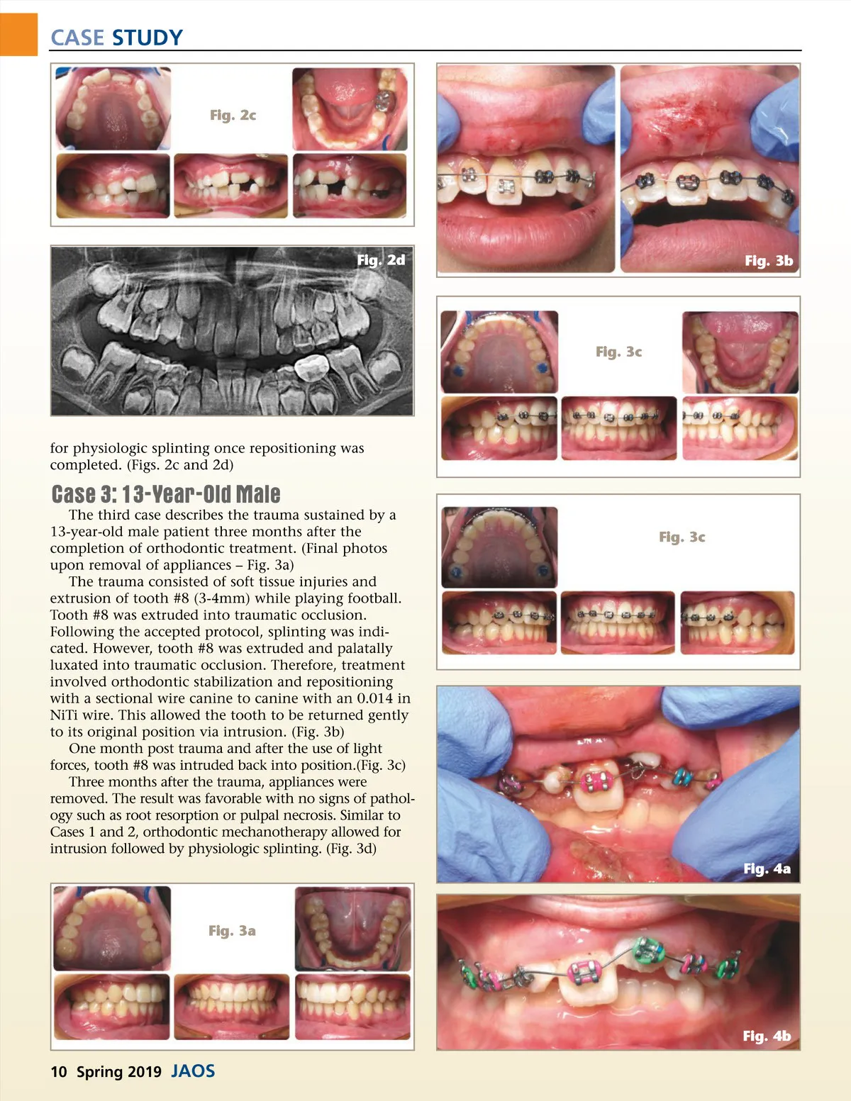
CASE STUDY Fig. 2c Fig. 2d Fig. 3b Fig. 3c for physiologic splinting once repositioning was completed. (Figs. 2c and 2d) Case 3: 13-Year-Old Male The third case describes the trauma sustained by a 13-year-old male patient three months after the completion of orthodontic treatment. (Final photos upon removal of appliances – Fig. 3a) The trauma consisted of soft tissue injuries and extrusion of tooth #8 (3-4mm) while playing football. Tooth #8 was extruded into traumatic occlusion. Following the accepted protocol, splinting was indi-cated. However, tooth #8 was extruded and palatally luxated into traumatic occlusion. Therefore, treatment involved orthodontic stabilization and repositioning with a sectional wire canine to canine with an 0.014 in NiTi wire. This allowed the tooth to be returned gently to its original position via intrusion. (Fig. 3b) One month post trauma and after the use of light forces, tooth #8 was intruded back into position.(Fig. 3c) Three months after the trauma, appliances were removed. The result was favorable with no signs of pathol-ogy such as root resorption or pulpal necrosis. Similar to Cases 1 and 2, orthodontic mechanotherapy allowed for intrusion followed by physiologic splinting. (Fig. 3d) Fig. 3c Fig. 4a Fig. 3a Fig. 4b 10 Spring 2019 JAOS
Journal of the American Orthodontic Society Spring 2019: Page 10