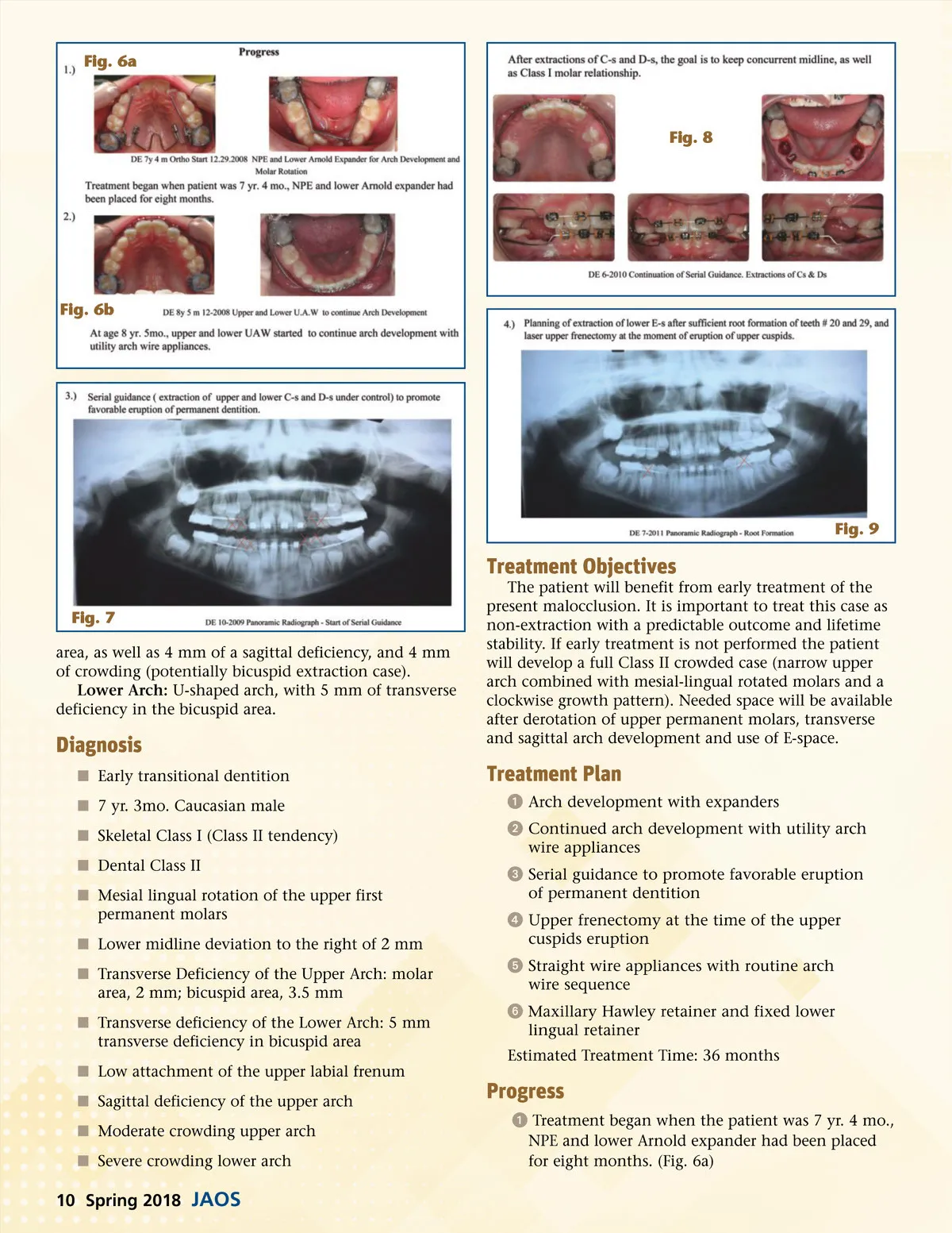
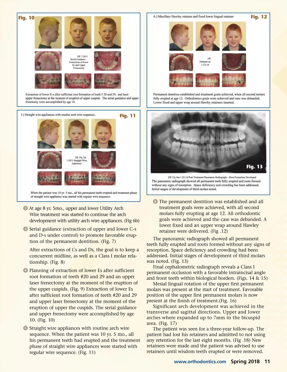
Fig. 10 Fig. 12 Fig. 11 Fig. 13 ᕢ At age 8 yr. 5mo., upper and lower Utility Arch Wire treatment was started to continue the arch development with utility arch wire appliances. (Fig 6b) ᕣ Serial guidance (extraction of upper and lower C-s and D-s under control) to promote favorable erup-tion of the permanent dentition. (Fig. 7) After extractions of Cs and Ds, the goal is to keep a concurrent midline, as well as a Class I molar rela-tionship. (Fig. 8) ᕤ Planning of extraction of lower Es after sufficient root formation of teeth #20 and 29 and an upper laser frenectomy at the moment of the eruption of the upper cuspids. (Fig. 9) Extraction of lower Es after sufficient root formation of teeth #20 and 29 and upper laser frenectomy at the moment of the eruption of upper the cuspids. The serial guidance and upper frenectomy were accomplished by age 10. (Fig. 10) ᕥ Straight wire appliances with routine arch wire sequence. When the patient was 10 yr. 5 mo., all his permanent teeth had erupted and the treatment phase of straight wire appliances were started with regular wire sequence. (Fig. 11) ᕦ The permanent dentition was established and all treatment goals were achieved, with all second molars fully erupting at age 12. All orthodontic goals were achieved and the case was debanded. A lower fixed and an upper wrap around Hawley retainer were delivered. (Fig .12) The panoramic radiograph showed all permanent teeth fully erupted and roots formed without any signs of resorption. Space deficiency and crowding had been addressed. Initial stages of development of third molars was noted. (Fig. 13) Final cephalometric radiograph reveals a Class I permanent occlusion with a favorable intraincisal angle and front teeth within biological borders. (Figs. 14 & 15) Mesial lingual rotation of the upper first permanent molars was present at the start of treatment. Favorable position of the upper first permanent molars is now present at the finish of treatment.(Fig. 16) Significant arch development was achieved in the transverse and sagittal directions. Upper and lower arches where expanded up to 7mm in the bicuspid area. (Fig. 17) The patient was seen for a three-year follow-up. The patient had lost his retainers and admitted to not using any retention for the last eight months. (Fig .18) New retainers were made and the patient was advised to use retainers until wisdom teeth erupted or were removed. www.orthodontics.com Spring 2018 11
Journal of the American Orthodontic Society Spring 2018: Page 11