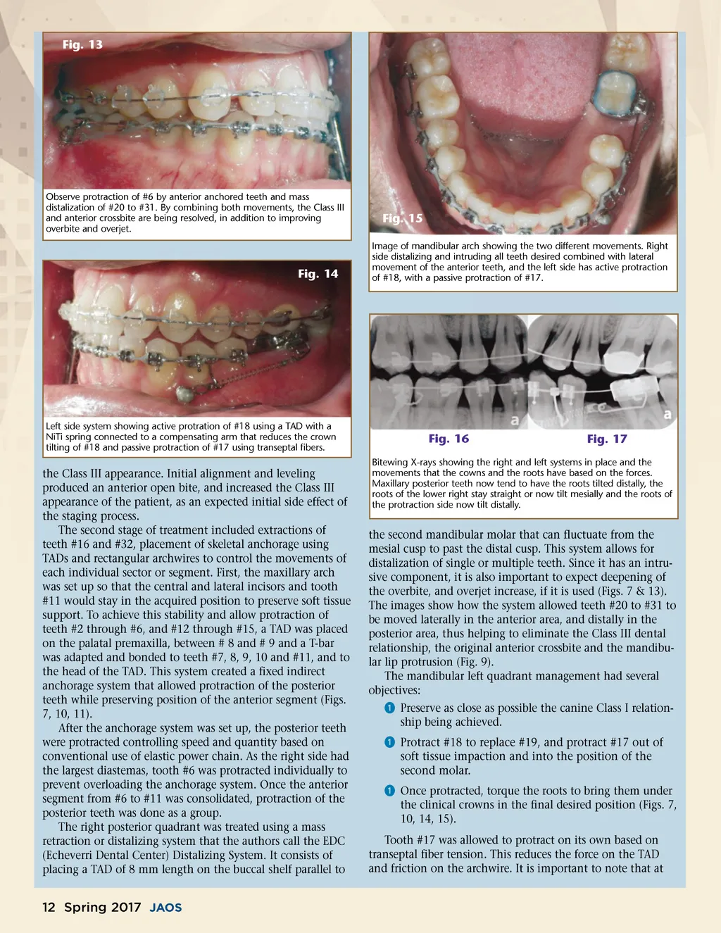
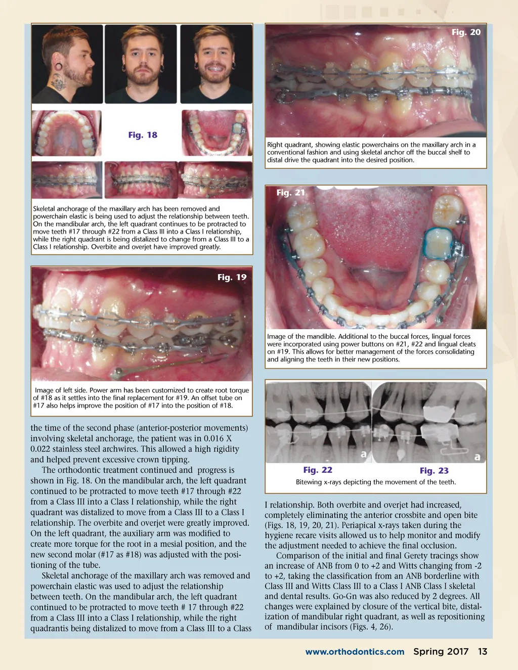
Fig. 20 Fig. 18 Right quadrant, showing elastic powerchains on the maxillary arch in a conventional fashion and using skeletal anchor off the buccal shelf to distal drive the quadrant into the desired position. Fig. 21 Skeletal anchorage of the maxillary arch has been removed and powerchain elastic is being used to adjust the relationship between teeth. On the mandibular arch, the left quadrant continues to be protracted to move teeth #17 through #22 from a Class III into a Class I relationship, while the right quadrant is being distalized to change from a Class III to a Class I relationship. Overbite and overjet have improved greatly. Fig. 19 Image of the mandible. Additional to the buccal forces, lingual forces were incorporated using power buttons on #21, #22 and lingual cleats on #19. This allows for better management of the forces consolidating and aligning the teeth in their new positions. Image of left side. Power arm has been customized to create root torque of #18 as it settles into the final replacement for #19. An offset tube on #17 also helps improve the position of #17 into the position of #18. the time of the second phase (anterior-posterior movements) involving skeletal anchorage, the patient was in 0.016 X 0.022 stainless steel archwires. This allowed a high rigidity and helped prevent excessive crown tipping. The orthodontic treatment continued and progress is shown in Fig. 18. On the mandibular arch, the left quadrant continued to be protracted to move teeth #17 through #22 from a Class III into a Class I relationship, while the right quadrant was distalized to move from a Class III to a Class I relationship. The overbite and overjet were greatly improved. On the left quadrant, the auxiliary arm was modified to create more torque for the root in a mesial position, and the new second molar (#17 as #18) was adjusted with the posi-tioning of the tube. Skeletal anchorage of the maxillary arch was removed and powerchain elastic was used to adjust the relationship between teeth. On the mandibular arch, the left quadrant continued to be protracted to move teeth # 17 through #22 from a Class III into a Class I relationship, while the right quadrantis being distalized to move from a Class III to a Class Fig. 22 Fig. 23 Bitewing x-rays depicting the movement of the teeth. I relationship. Both overbite and overjet had increased, completely eliminating the anterior crossbite and open bite (Figs. 18, 19, 20, 21). Periapical x-rays taken during the hygiene recare visits allowed us to help monitor and modify the adjustment needed to achieve the final occlusion. Comparison of the initial and final Gerety tracings show an increase of ANB from 0 to +2 and Witts changing from -2 to +2, taking the classification from an ANB borderline with Class III and Witts Class III to a Class I ANB Class I skeletal and dental results. Go-Gn was also reduced by 2 degrees. All changes were explained by closure of the vertical bite, distal-ization of mandibular right quadrant, as well as repositioning of mandibular incisors (Figs. 4, 26). www.orthodontics.com Spring 2017 13
Journal of the American Orthodontic Society Spring 2017: Page 13