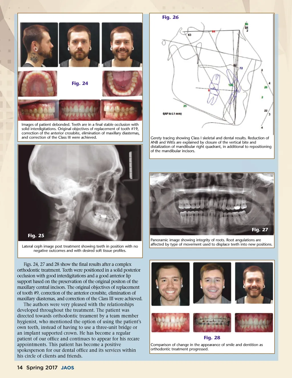
Fig. 26 Fig. 24 Images of patient debonded. Teeth are in a final stable occlusion with solid interdigitations. Original objectives of replacement of tooth #19, correction of the anterior crossbite, elimination of maxillary diastemas, and correction of the Class III were achieved. Gerety tracing showing Class I skeletal and dental results. Reduction of ANB and Witts are explained by closure of the vertical bite and distalization of mandibular right quadrant, in additional to repositioning of the mandibular incisors. Fig. 2 7 Fig. 2 5 Lateral ceph image post treatment showing teeth in position with no negative outcomes and with desired soft tissue profiles. Panoramic image showing integrity of roots. Root angulations are affected by type of movement used to displace teeth into new positions. Figs. 24, 27 and 28 show the final results after a complex orthodontic treatment. Teeth were positioned in a solid posterior occlusion with good interdigitations and a good anterior lip support based on the preservation of the original positon of the maxillary central incisors. The original objectives of replacement of tooth #9, correction of the anterior crossbite, elimination of maxillary diastemas, and correction of the Class III were achieved. The authors were very pleased with the relationships developed throughout the treatment. The patient was directed towards orthodontic treament by a team member hygienist, who mentioned the option of using the patient’s own teeth, instead of having to use a three-unit bridge or an implant supported crown. He has become a regular patient of our office and continues to appear for his recare appointments. This patient has become a positive spokesperson for our dental office and its services within his circle of clients and friends. Fig. 28 Comparison of change in the appearance of smile and dentition as orthodontic treatment progressed. 14 Spring 2017 JAOS
Journal of the American Orthodontic Society Spring 2017: Page 14