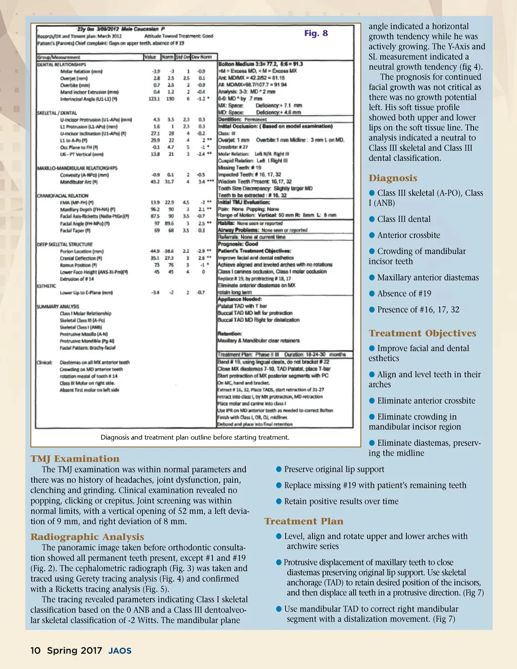
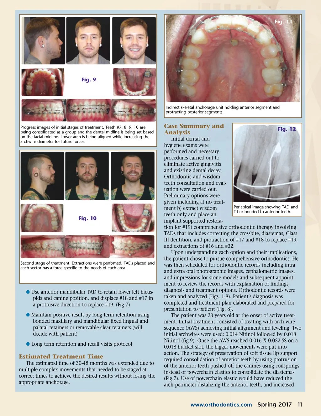
Fig. 11 Fig. 9 Indirect skeletal anchorage unit holding anterior segment and protracting posterior segments. Progress images of initial stages of treatment. Teeth #7, 8, 9, 10 are being consolidated as a group and the dental midline is being set based on the facial midline. Lower arch is being aligned while increasing the archwire diameter for future forces. Case Summary and Analysis Fig. 12 Fig. 10 Second stage of treatment. Extractions were perfomed, TADs placed and each sector has a force specific to the needs of each area. b Use anterior mandibular TAD to retain lower left bicus-pids and canine position, and displace #18 and #17 in a protrusive direction to replace #19. (Fig 7) b Maintain positive result by long term retention using bonded maxillary and mandibular fixed lingual and palatal retainers or removable clear retainers (will decide with patient) b Long term retention and recall visits protocol Estimated Treatment Time The estimated time of 30-48 months was extended due to multiple complex movements that needed to be staged at correct times to achieve the desired results without losing the appropriate anchorage. Initial dental and hygiene exams were performed and necessary procedures carried out to eliminate active gingivitis and existing dental decay. Orthodontic and wisdom teeth consultation and eval-uation were carried out. Preliminary options were given including a) no treat-Periapical image showing TAD and ment b) extract wisdom T-bar bonded to anterior teeth. teeth only and place an implant supported restora-tion for #19) comprehensive orthodontic therapy involving TADs that includes correcting the crossbite, diastemas, Class III dentition, and protraction of #17 and #18 to replace #19, and extractions of #16 and #32. Upon understanding each option and their implications, the patient chose to pursue comprehensive orthodontics. He was then scheduled for orthodontic records including intra and extra oral photographic images, cephalometric images, and impressions for stone models and subsequent appoint-ment to review the records with explanation of findings, diagnosis and treatment options. Orthodontic records were taken and analyzed (Figs. 1-8). Patient’s diagnosis was completed and treatment plan elaborated and prepared for presentation to patient (Fig. 8). The patient was 23 years old at the onset of active treat-ment. Initial treatment consisted of treating with arch wire sequence (AWS) achieving initial alignment and leveling. Two initial archwires were used; 0.014 Nitinol followed by 0.018 Nitinol (fig 9). Once the AWS reached 0.016 X 0.022 SS on a 0.018 bracket slot, the bigger movements were put into action. The strategy of preservation of soft tissue lip support required consolidation of anterior teeth by using protrusion of the anterior teeth pushed off the canines using coilsprings instead of powerchain elastics to consolidate the diastemas (Fig 7). Use of powerchain elastic would have reduced the arch perimeter distalizing the anterior teeth, and increased www.orthodontics.com Spring 2017 11
Journal of the American Orthodontic Society Spring 2017: Page 11