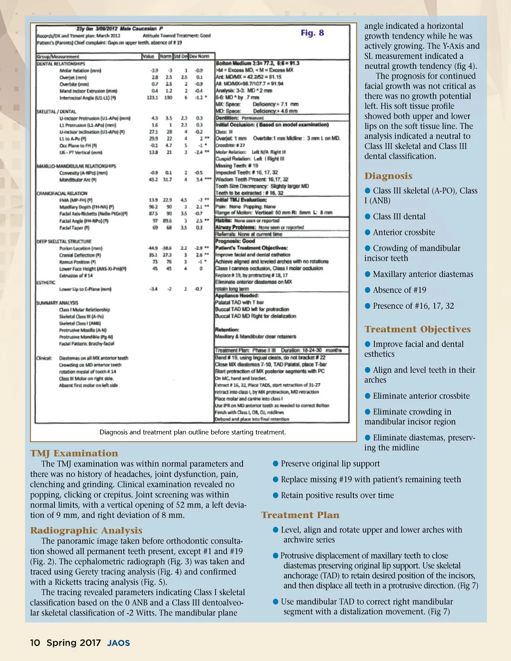
Fig. 8 angle indicated a horizontal growth tendency while he was actively growing. The Y-Axis and SL measurement indicated a neutral growth tendency (fig 4). The prognosis for continued facial growth was not critical as there was no growth potential left. His soft tissue profile showed both upper and lower lips on the soft tissue line. The analysis indicated a neutral to Class III skeletal and Class III dental classification. Diagnosis b Class III skeletal (A-PO), Class I (ANB) b Class III dental b Anterior crossbite b Crowding of mandibular incisor teeth b Maxillary anterior diastemas b Absence of #19 b Presence of #16, 17, 32 Treatment Objectives b Improve facial and dental esthetics b Align and level teeth in their arches b Eliminate anterior crossbite b Eliminate crowding in mandibular incisor region Diagnosis and treatment plan outline before starting treatment. TMJ Examination The TMJ examination was within normal parameters and there was no history of headaches, joint dysfunction, pain, clenching and grinding. Clinical examination revealed no popping, clicking or crepitus. Joint screening was within normal limits, with a vertical opening of 52 mm, a left devia-tion of 9 mm, and right deviation of 8 mm. b Eliminate diastemas, preserv-ing the midline b Preserve original lip support b Replace missing #19 with patient’s remaining teeth b Retain positive results over time Treatment Plan b Level, align and rotate upper and lower arches with archwire series b Protrusive displacement of maxillary teeth to close diastemas preserving original lip support. Use skeletal anchorage (TAD) to retain desired position of the incisors, and then displace all teeth in a protrusive direction. (Fig 7) b Use mandibular TAD to correct right mandibular segment with a distalization movement. (Fig 7) Radiographic Analysis The panoramic image taken before orthodontic consulta-tion showed all permanent teeth present, except #1 and #19 (Fig. 2). The cephalometric radiograph (Fig. 3) was taken and traced using Gerety tracing analysis (Fig. 4) and confirmed with a Ricketts tracing analysis (Fig. 5). The tracing revealed parameters indicating Class I skeletal classification based on the 0 ANB and a Class III dentoalveo-lar skeletal classification of -2 Witts. The mandibular plane 10 Spring 2017 JAOS
Journal of the American Orthodontic Society Spring 2017: Page 10