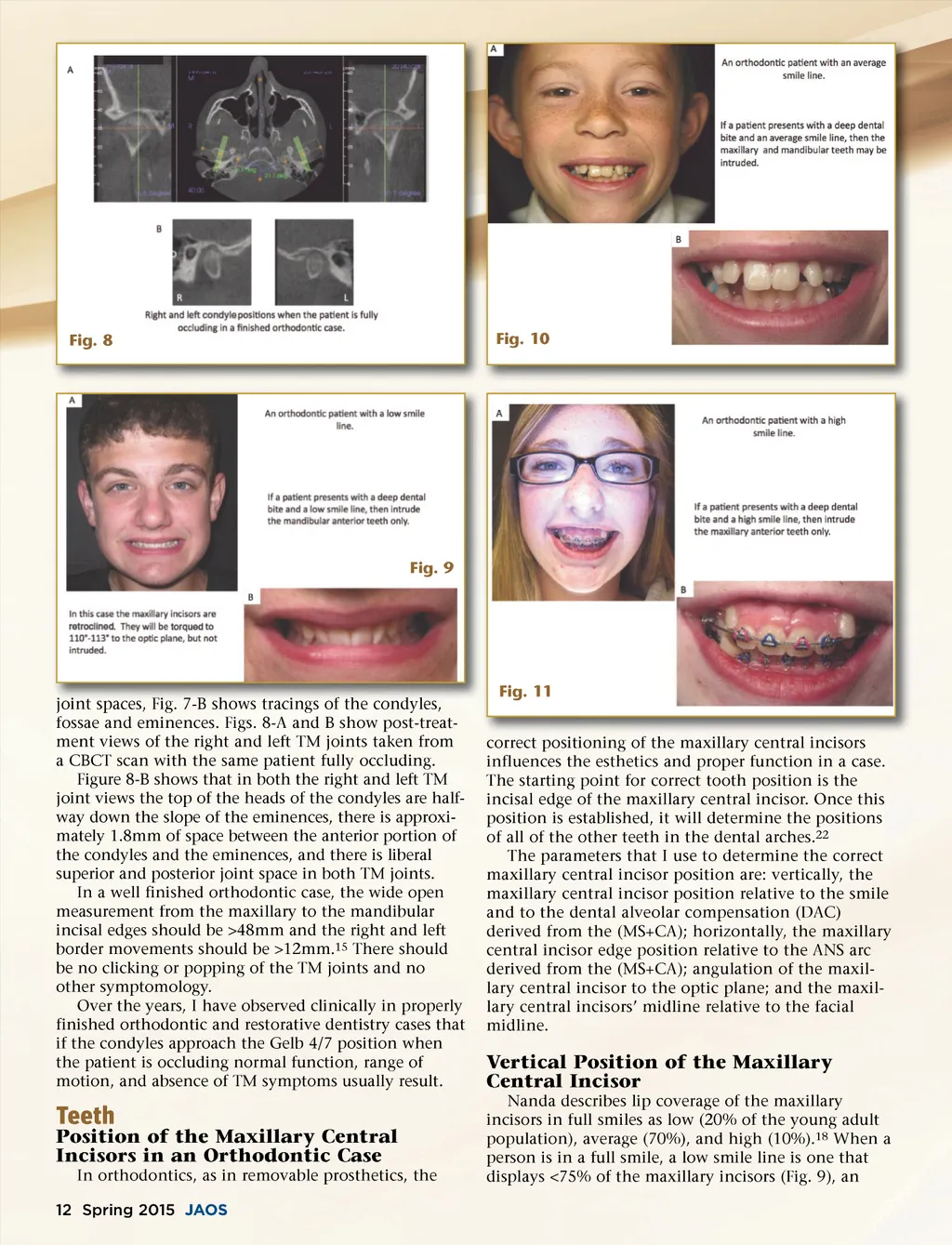
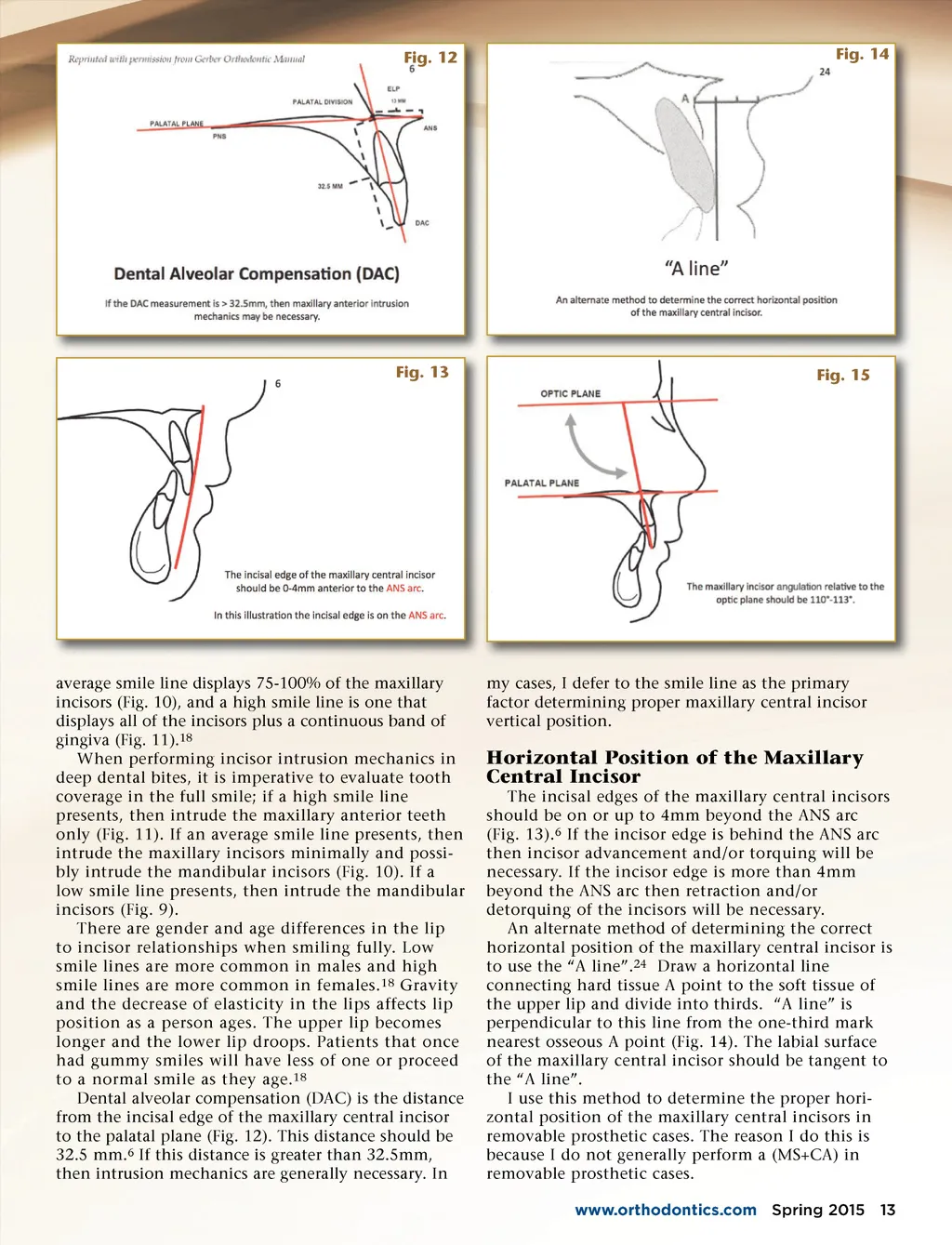
Fig. 8 Fig. 10 Fig. 9 joint spaces, spaces Fig. Fig 7-B 7 B shows tracings of the condyles, condyles fossae and eminences. Figs. 8-A and B show post-treat-ment views of the right and left TM joints taken from a CBCT scan with the same patient fully occluding. Figure 8-B shows that in both the right and left TM joint views the top of the heads of the condyles are half-way down the slope of the eminences, there is approxi-mately 1.8mm of space between the anterior portion of the condyles and the eminences, and there is liberal superior and posterior joint space in both TM joints. In a well finished orthodontic case, the wide open measurement from the maxillary to the mandibular incisal edges should be >48mm and the right and left border movements should be >12mm. 15 There should be no clicking or popping of the TM joints and no other symptomology. Over the years, I have observed clinically in properly finished orthodontic and restorative dentistry cases that if the condyles approach the Gelb 4/7 position when the patient is occluding normal function, range of motion, and absence of TM symptoms usually result. Fig. 11 correct positioning of central incisors ii i f the h maxillary ill li i influences the esthetics and proper function in a case. The starting point for correct tooth position is the incisal edge of the maxillary central incisor. Once this position is established, it will determine the positions of all of the other teeth in the dental arches. 22 The parameters that I use to determine the correct maxillary central incisor position are: vertically, the maxillary central incisor position relative to the smile and to the dental alveolar compensation (DAC) derived from the (MS+CA); horizontally, the maxillary central incisor edge position relative to the ANS arc derived from the (MS+CA); angulation of the maxil-lary central incisor to the optic plane; and the maxil-lary central incisors’ midline relative to the facial midline. Vertical Position of the Maxillary Central Incisor Nanda describes lip coverage of the maxillary incisors in full smiles as low (20% of the young adult population), average (70%), and high (10%). 18 When a person is in a full smile, a low smile line is one that displays <75% of the maxillary incisors (Fig. 9), an Teeth Position of the Maxillary Central Incisors in an Orthodontic Case In orthodontics, as in removable prosthetics, the 12 Spring 2015 JAOS
Journal of the American Orthodontic Society Spring 2015: Page 12