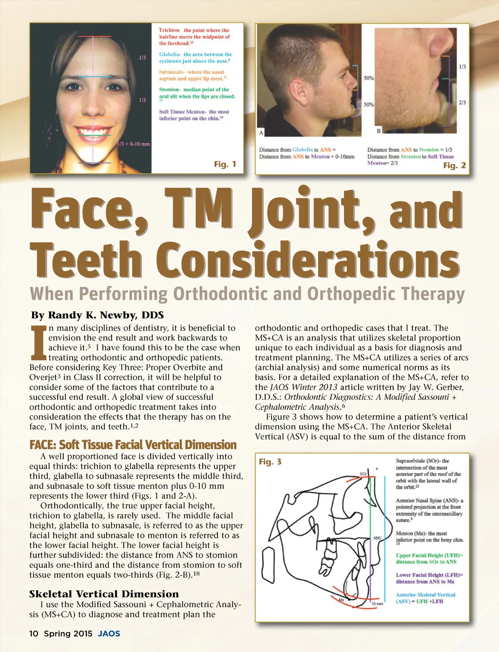
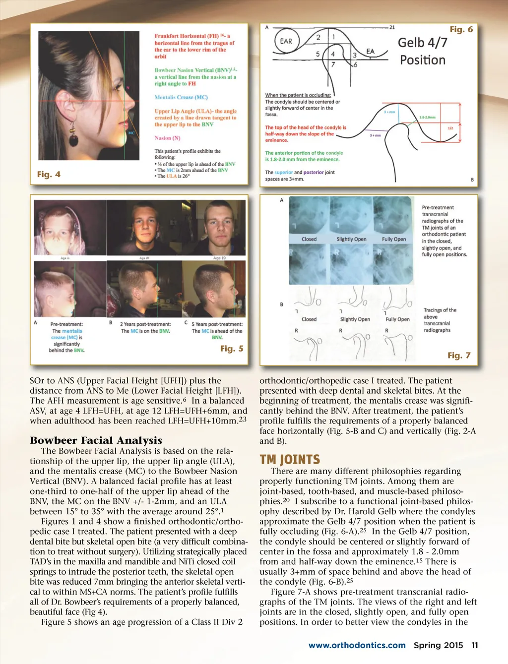
Fig. 6 Fig. 4 Fig. 5 Fig. 7 orthodontic/orthopedic case I treated. The patient presented with deep dental and skeletal bites. At the beginning of treatment, the mentalis crease was signifi-cantly behind the BNV. After treatment, the patient’s profile fulfills the requirements of a properly balanced face horizontally (Fig. 5-B and C) and vertically (Fig. 2-A and B). SOr to ANS (Upper Facial Height [UFH]) plus the distance from ANS to Me (Lower Facial Height [LFH]). The AFH measurement is age sensitive. 6 In a balanced ASV, at age 4 LFH=UFH, at age 12 LFH=UFH+6mm, and when adulthood has been reached LFH=UFH+10mm. 23 Bowbeer Facial Analysis The Bowbeer Facial Analysis is based on the rela-tionship of the upper lip, the upper lip angle (ULA), and the mentalis crease (MC) to the Bowbeer Nasion Vertical (BNV). A balanced facial profile has at least one-third to one-half of the upper lip ahead of the BNV, the MC on the BNV +/-1-2mm, and an ULA between 15° to 35° with the average around 25°. 1 Figures 1 and 4 show a finished orthodontic/ortho-pedic case I treated. The patient presented with a deep dental bite but skeletal open bite (a very difficult combina-tion to treat without surgery). Utilizing strategically placed TAD’s in the maxilla and mandible and NiTi closed coil springs to intrude the posterior teeth, the skeletal open bite was reduced 7mm bringing the anterior skeletal verti-cal to within MS+CA norms. The patient’s profile fulfills all of Dr. Bowbeer’s requirements of a properly balanced, beautiful face (Fig 4). Figure 5 shows an age progression of a Class II Div 2 TM JOINTS There are many different philosophies regarding properly functioning TM joints. Among them are joint-based, tooth-based, and muscle-based philoso-phies. 20 I subscribe to a functional joint-based philos-ophy described by Dr. Harold Gelb where the condyles approximate the Gelb 4/7 position when the patient is fully occluding (Fig. 6-A). 25 In the Gelb 4/7 position, the condyle should be centered or slightly forward of center in the fossa and approximately 1.8 -2.0mm from and half-way down the eminence. 15 There is usually 3+mm of space behind and above the head of the condyle (Fig. 6-B). 25 Figure 7-A shows pre-treatment transcranial radio-graphs of the TM joints. The views of the right and left joints are in the closed, slightly open, and fully open positions. In order to better view the condyles in the www.orthodontics.com Spring 2015 11
Journal of the American Orthodontic Society Spring 2015: Page 11