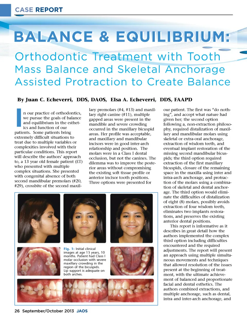
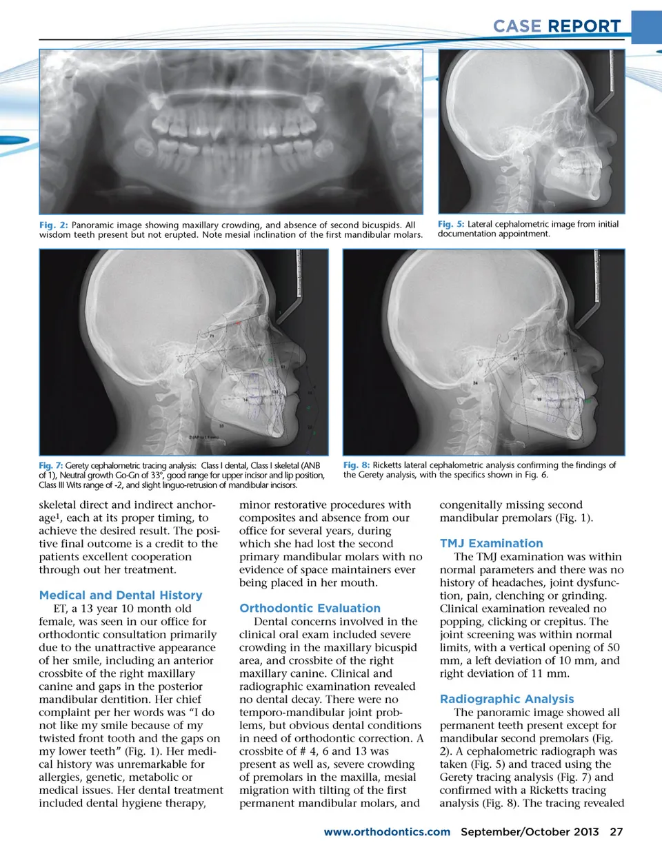
CASE REPORT Fig. 2: Panoramic image showing maxillary crowding, and absence of second bicuspids. All wisdom teeth present but not erupted. Note mesial inclination of the first mandibular molars. Fig. 5: Lateral cephalometric image from initial documentation appointment. Fig. 7: Gerety cephalometric tracing analysis: Class I dental, Class I skeletal (ANB of 1), Neutral growth Go-Gn of 33°, good range for upper incisor and lip position, Class III Wits range of -2, and slight linguo-retrusion of mandibular incisors. Fig. 8: Ricketts lateral cephalometric analysis confirming the findings of the Gerety analysis, with the specifics shown in Fig. 6. skeletal direct and indirect anchor-age 1 , each at its proper timing, to achieve the desired result. The posi-tive final outcome is a credit to the patients excellent cooperation through out her treatment. minor restorative procedures with composites and absence from our office for several years, during which she had lost the second primary mandibular molars with no evidence of space maintainers ever being placed in her mouth. congenitally missing second mandibular premolars (Fig. 1). TMJ Examination The TMJ examination was within normal parameters and there was no history of headaches, joint dysfunc-tion, pain, clenching or grinding. Clinical examination revealed no popping, clicking or crepitus. The joint screening was within normal limits, with a vertical opening of 50 mm, a left deviation of 10 mm, and right deviation of 11 mm. Medical and Dental History ET, a 13 year 10 month old female, was seen in our office for orthodontic consultation primarily due to the unattractive appearance of her smile, including an anterior crossbite of the right maxillary canine and gaps in the posterior mandibular dentition. Her chief complaint per her words was “I do not like my smile because of my twisted front tooth and the gaps on my lower teeth” (Fig. 1). Her medi-cal history was unremarkable for allergies, genetic, metabolic or medical issues. Her dental treatment included dental hygiene therapy, Orthodontic Evaluation Dental concerns involved in the clinical oral exam included severe crowding in the maxillary bicuspid area, and crossbite of the right maxillary canine. Clinical and radiographic examination revealed no dental decay. There were no temporo-mandibular joint prob-lems, but obvious dental conditions in need of orthodontic correction. A crossbite of # 4, 6 and 13 was present as well as, severe crowding of premolars in the maxilla, mesial migration with tilting of the first permanent mandibular molars, and Radiographic Analysis The panoramic image showed all permanent teeth present except for mandibular second premolars (Fig. 2). A cephalometric radiograph was taken (Fig. 5) and traced using the Gerety tracing analysis (Fig. 7) and confirmed with a Ricketts tracing analysis (Fig. 8). The tracing revealed www.orthodontics.com September/October 2013 27
Journal of the American Orthodontic Society September-October 2013: Page 27