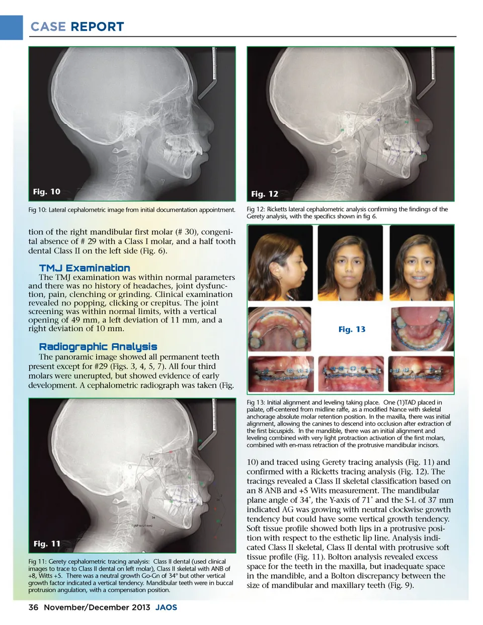
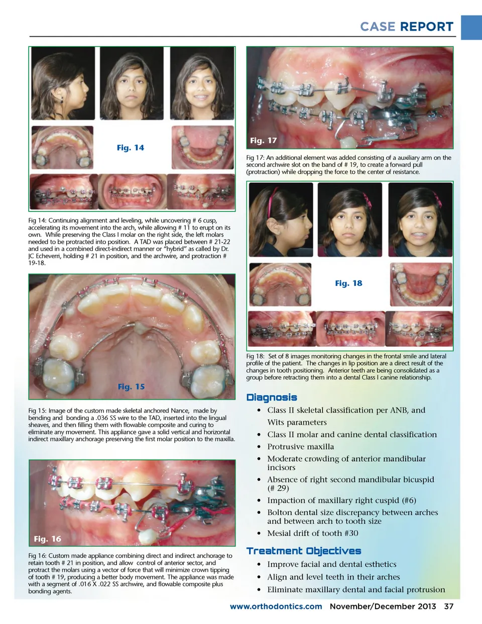
CASE REPORT Fig. 10 Fig 10: Lateral cephalometric image from initial documentation appointment. Fig. 12 Fig 12: Ricketts lateral cephalometric analysis confirming the findings of the Gerety analysis, with the specifics shown in fig 6. tion of the right mandibular first molar (# 30), congeni-tal absence of # 29 with a Class I molar, and a half tooth dental Class II on the left side (Fig. 6). f; $a;#"$ #c;" The TMJ examination was within normal parameters and there was no history of headaches, joint dysfunc-tion, pain, clenching or grinding. Clinical examination revealed no popping, clicking or crepitus. The joint screening was within normal limits, with a vertical opening of 49 mm, a left deviation of 11 mm, and a right deviation of 10 mm. Fig. 13 $#c;�f;d;$�e;#f;�b;"$b;e;#e; The panoramic image showed all permanent teeth present except for #29 (Figs. 3, 4, 5, 7). All four third molars were unerupted, but showed evidence of early development. A cephalometric radiograph was taken (Fig. Fig 13: Initial alignment and leveling taking place. One (1)TAD placed in palate, off-centered from midline raffe, as a modified Nance with skeletal anchorage absolute molar retention position. In the maxilla, there was initial alignment, allowing the canines to descend into occlusion after extraction of the first bicuspids. In the mandible, there was an initial alignment and leveling combined with very light protraction activation of the first molars, combined with en-mass retraction of the protrusive mandibular incisors. Fig. 11 Fig 11: Gerety cephalometric tracing analysis: Class II dental (used clinical images to trace to Class II dental on left molar), Class II skeletal with ANB of +8, Witts +5. There was a neutral growth Go-Gn of 34° but other vertical growth factor indicated a vertical tendency. Mandibular teeth were in buccal protrusion angulation, with a compensation position. 10) and traced using Gerety tracing analysis (Fig. 11) and confirmed with a Ricketts tracing analysis (Fig. 12). The tracings revealed a Class II skeletal classification based on an 8 ANB and +5 Wits measurement. The mandibular plane angle of 34˚, the Y-axis of 71˚ and the S-L of 37 mm indicated AG was growing with neutral clockwise growth tendency but could have some vertical growth tendency. Soft tissue profile showed both lips in a protrusive posi-tion with respect to the esthetic lip line. Analysis indi-cated Class II skeletal, Class II dental with protrusive soft tissue profile (Fig. 11). Bolton analysis revealed excess space for the teeth in the maxilla, but inadequate space in the mandible, and a Bolton discrepancy between the size of mandibular and maxillary teeth (Fig. 9). 36 November/December 2013 JAOS
Journal of the American Orthodontic Society November-December 2013: Page 36