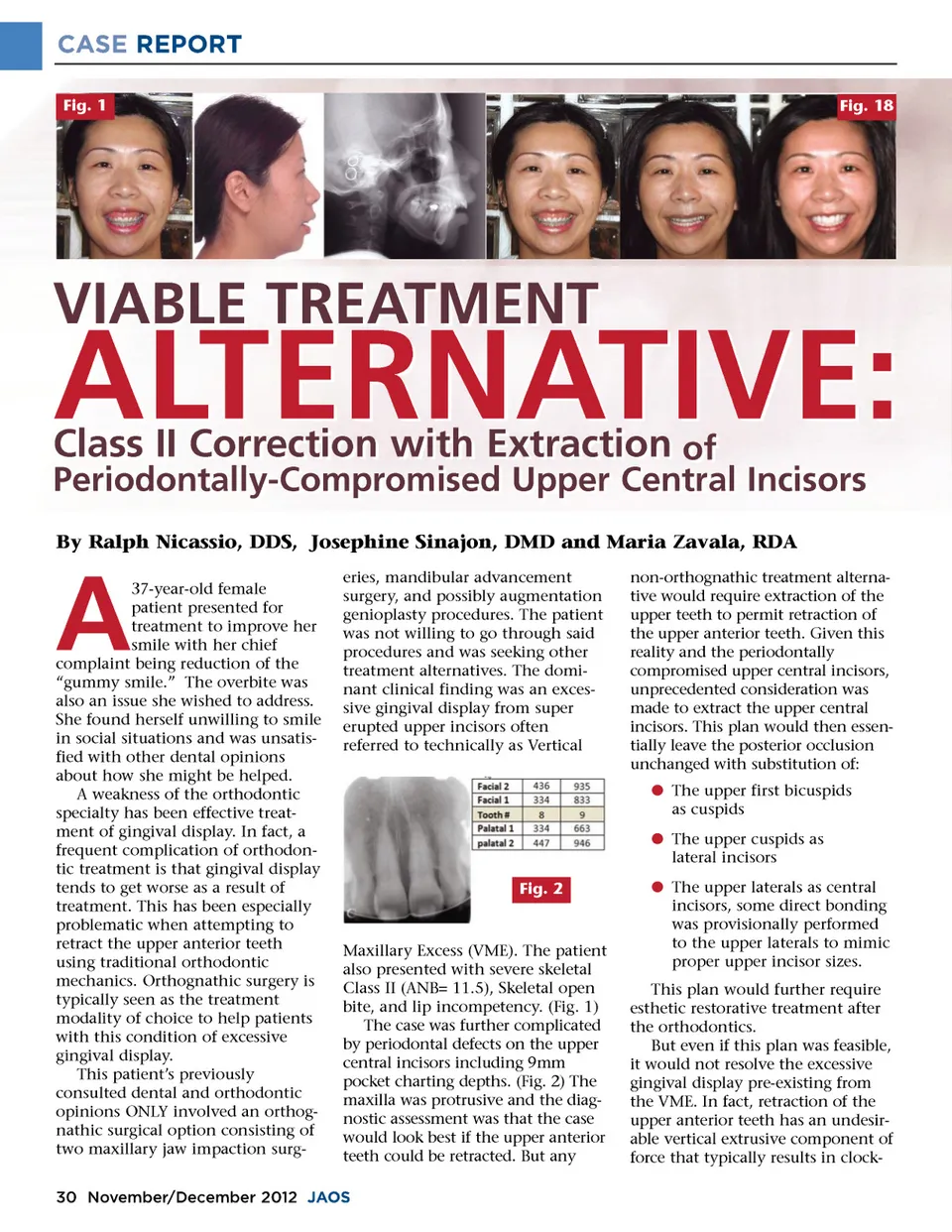
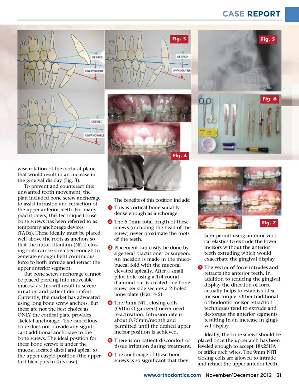
CASE REPORT Fig. 3 Fig. 5 Fig. 6 Fig. 4 wise rotation of the occlusal plane that would result in an increase in the gingival display (Fig. 3). To prevent and counteract this unwanted tooth movement, the plan included bone screw anchorage to assist intrusion and retraction of the upper anterior teeth. For many practitioners, this technique to use bone screws has been referred to as temporary anchorage devices (TADs). These ideally must be placed well above the roots as anchors so that the nickel titanium (NiTi) clos-ing coils can be stretched enough to generate enough light continuous force to both intrude and retract the upper anterior segment. But bone screw anchorage cannot be placed piercing into moveable mucosa as this will result in severe irritation and patient discomfort. Currently, the market has advocated using long bone screw anchors. But these are not the best choice as ONLY the cortical plate provides skeletal anchorage. The cancellous bone does not provide any signifi-cant additional anchorage to the bone screws. The ideal position for these bone screws is under the mucosa located distal and apical to the upper cuspid position (the upper first bicuspids in this case). The benefits of this position include: ᕡ This is cortical bone suitably dense enough as anchorage. ᕢ The 6.0mm total length of these screws (including the head of the screw) never proximate the roots of the teeth. ᕣ Placement can easily be done by a general practitioner or surgeon. An incision is made in the muco-buccal fold with the mucosal elevated apically. After a small pilot hole using a 1/4 round diamond bur is created one bone screw per side secures a 2-holed bone plate (Figs. 4-5). ᕤ The 9mm NiTi closing coils (Ortho Organizers) never need re-activation. Intrusion rate is about 0.75mm/month and permitted until the desired upper incisor position is achieved. ᕥ There is no patient discomfort or tissue irritation during treatment. ᕦ The anchorage of these bone screws is so significant that they Fig. 7 later permit using anterior verti-cal elastics to extrude the lower incisors without the anterior teeth extruding which would exacerbate the gingival display. ᕧ The vector of force intrudes and retracts the anterior teeth. In addition to reducing the gingival display the direction of force actually helps to establish ideal incisor torque. Other traditional orthodontic incisor retraction techniques tend to extrude and de-torque the anterior segments resulting in an increase in gingi-val display. Ideally, the bone screws should be placed once the upper arch has been leveled enough to accept 18x25HA or stiffer arch wires. The 9mm NiTi closing coils are allowed to intrude and retract the upper anterior teeth www.orthodontics.com November/December 2012 31
Journal of the American Orthodontic Society November-December 2012: Page 31