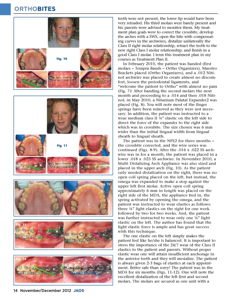
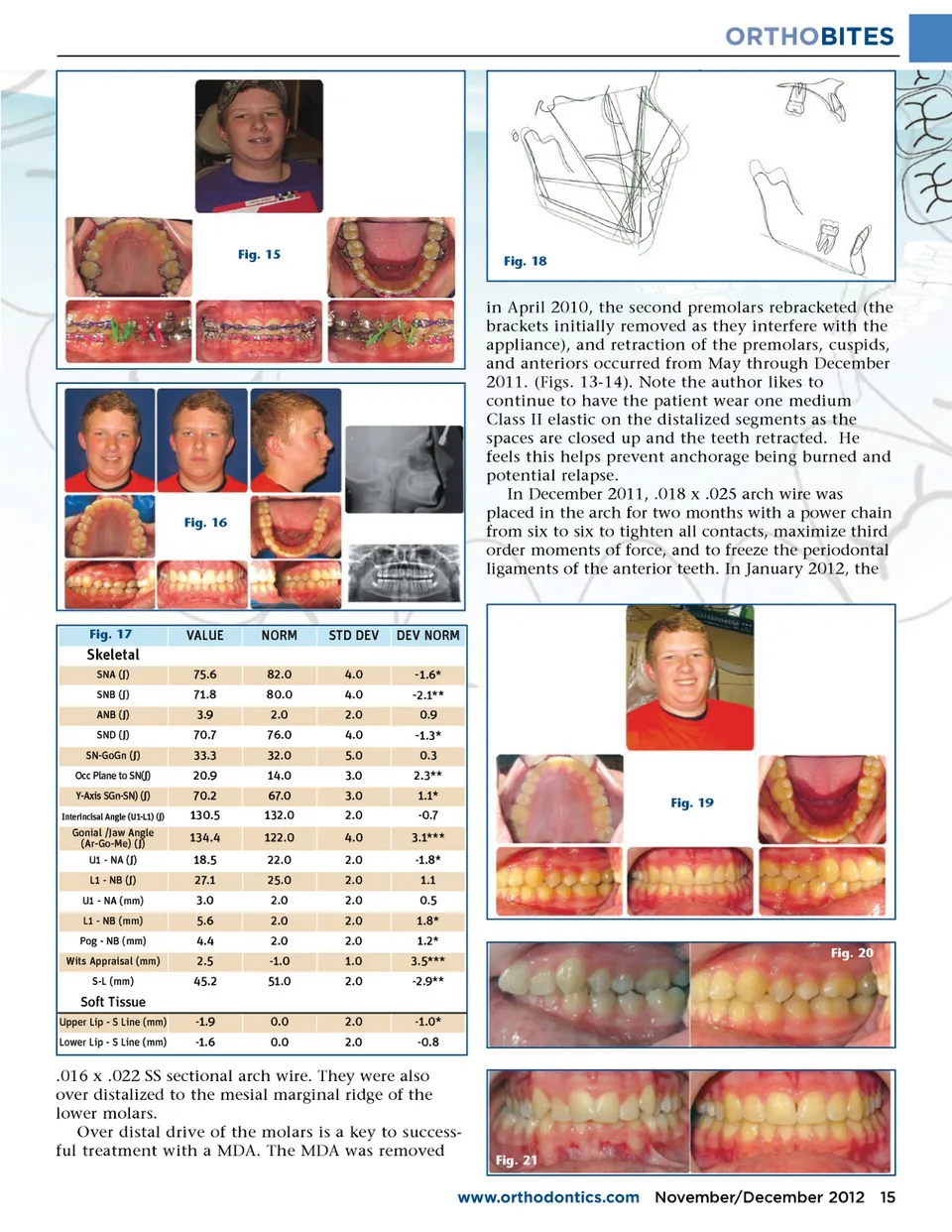
ORTHOBITES Fig. 15 Fig. 18 Fig. 16 in April 2010, the second premolars rebracketed (the brackets initially removed as they interfere with the appliance), and retraction of the premolars, cuspids, and anteriors occurred from May through December 2011. (Figs. 13-14). Note the author likes to continue to have the patient wear one medium Class II elastic on the distalized segments as the spaces are closed up and the teeth retracted. He feels this helps prevent anchorage being burned and potential relapse. In December 2011, .018 x .025 arch wire was placed in the arch for two months with a power chain from six to six to tighten all contacts, maximize third order moments of force, and to freeze the periodontal ligaments of the anterior teeth. In January 2012, the Fig. 17 VALUE 75.6 71.8 3.9 70.7 33.3 20.9 70.2 130.5 134.4 18.5 27.1 3.0 5.6 4.4 2.5 45.2 -1.9 -1.6 NORM 82.0 80.0 2.0 76.0 32.0 14.0 67.0 132.0 122.0 22.0 25.0 2.0 2.0 2.0 -1.0 51.0 0.0 0.0 STD DEV 4.0 4.0 2.0 4.0 5.0 3.0 3.0 2.0 4.0 2.0 2.0 2.0 2.0 2.0 1.0 2.0 2.0 2.0 DEV NORM -1.6* -2.1** 0.9 Skeletal SNA ( ∫ ) SNB ( ∫ ) ANB ( ∫ ) SND ( ∫ ) SN-GoGn ( ∫ ) Occ Plane to SN( ∫ ) Y-Axis SGn-SN) ( ∫ ) Interincisal Angle (U1-L1) ( ∫ ) -1.3* 0.3 2.3** 1.1* -0.7 3.1*** -1.8* 1.1 0.5 1.8* 1.2* 3.5*** -2.9** -1.0* -0.8 Fig. 20 Fig. 19 Gonial /Jaw Angle (Ar-Go-Me) ( ∫ ) U1 -NA ( ∫ ) L1 -NB ( ∫ ) U1 -NA (mm) L1 -NB (mm) Pog -NB (mm) Wits Appraisal (mm) S-L (mm) Soft Tissue Upper Lip -S Line (mm) Lower Lip -S Line (mm) .016 x .022 SS sectional arch wire. They were also over distalized to the mesial marginal ridge of the lower molars. Over distal drive of the molars is a key to success-ful treatment with a MDA. The MDA was removed Fig. 21 www.orthodontics.com November/December 2012 15
Journal of the American Orthodontic Society November-December 2012: Page 15