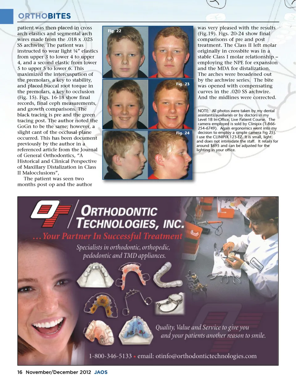
ORTHOBITES patient was then placed in cross arch elastics and segmental arch wires made from the .018 x .025 SS archwire. The patient was instructed to wear light ¼” elastics from upper 3 to lower 4 to upper 4, and a second elastic from lower 5 to upper 5 to lower 6. This maximized the intercuspation of the premolars, a key to stability, and placed buccal root torque in the premolars, a key to occlusion (Fig. 15). Figs. 16-18 show final records, final ceph measurements, and growth comparisons. The black tracing is pre and the green tracing post. The author noted the GoGn to be the same; however, a slight cant of the occlusal plane occurred. This has been document previously by the author in a referenced article from the Journal of General Orthodontics, “A Historical and Clinical Perspective of Maxillary Distalization in Class II Malocclusions”, The patient was seen two months post op and the author Fig. 22 Fig. 23 was very pleased with the results w (Fig.19). Figs. 20-24 show final ( comparisons of pre and post c treatment. The Class II left molar t originally in crossbite was in a o stable Class I molar relationship – s employing the NPE for expansion e and the MDA for distalization. a The arches were broadened out T by the archwire series. The bite b was opened with compensating w curves in the .020 SS archwire. c And the midlines were corrected. A N NOTE: All photos were taken by my dental a assistants/auxiliaries or by doctors in my L Level 1B In-Office, Live Patient Course. The c camera employed is sold by Clinipix (1-866-2 254-6749). Again ergonomics went into my d decision to employ a simple camera Fig 25). I use the CLINIPIX 123-EZ. It is small, light a and does not intimidate the staff. It retails for a around $695 and can be adjusted for the li lighting in your office. Fig. 24 16 November/December 2012 JAOS
Journal of the American Orthodontic Society November-December 2012: Page 16