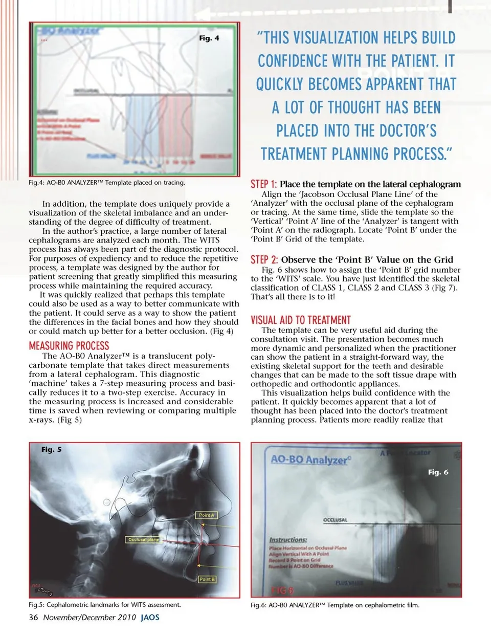
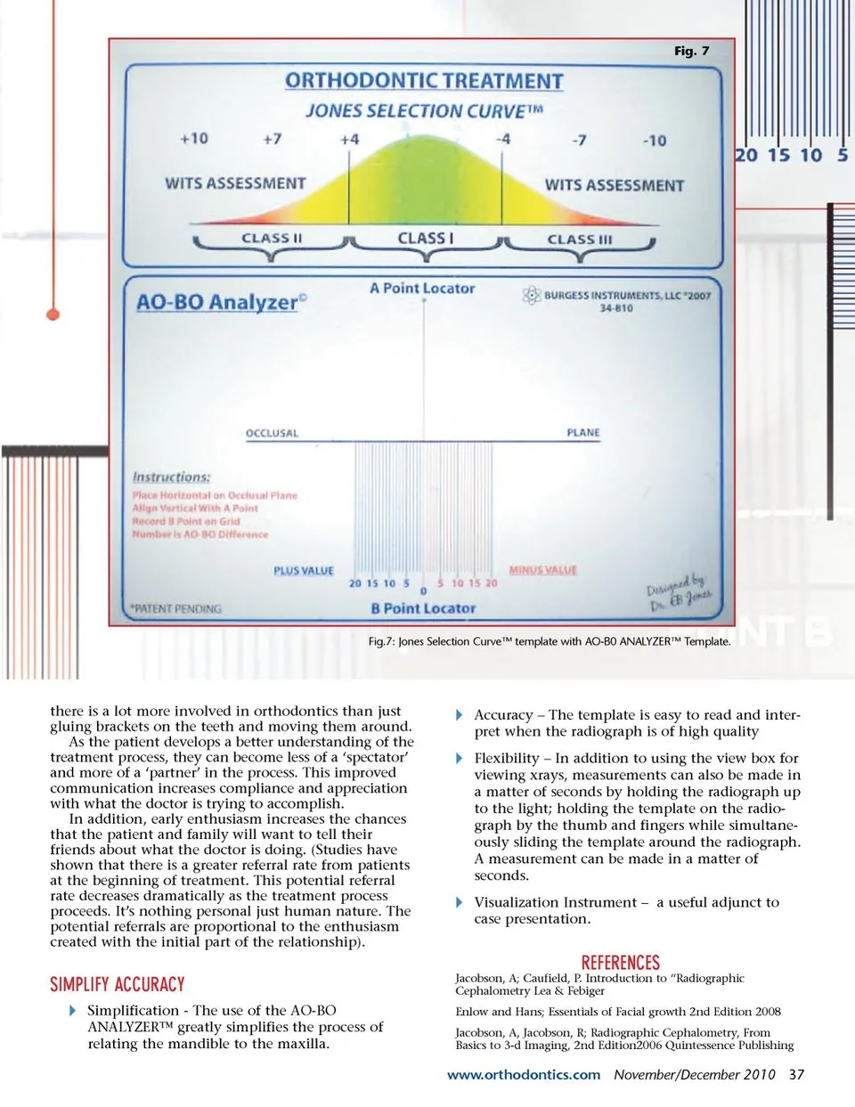
Fig. 4 “THIS VISUALIZATION HELPS BUILD CONFIDENCE WITH THE PATIENT. IT QUICKLY BECOMES APPARENT THAT A LOT OF THOUGHT HAS BEEN PLACED INTO THE DOCTOR’S TREATMENT PLANNING PROCESS.” STEP 1: Place the template on the lateral cephalogram Align the ‘Jacobson Occlusal Plane Line’ of the ‘Analyzer’ with the occlusal plane of the cephalogram or tracing. At the same time, slide the template so the ‘Vertical’ ‘Point A’ line of the ‘Analyzer’ is tangent with ‘Point A’ on the radiograph. Locate ‘Point B’ under the ‘Point B’ Grid of the template. Fig.4: AO-B0 ANALYZER™ Template placed on tracing. In addition, the template does uniquely provide a visualization of the skeletal imbalance and an under-standing of the degree of difficulty of treatment. In the author’s practice, a large number of lateral cephalograms are analyzed each month. The WITS process has always been part of the diagnostic protocol. For purposes of expediency and to reduce the repetitive process, a template was designed by the author for patient screening that greatly simplified this measuring process while maintaining the required accuracy. It was quickly realized that perhaps this template could also be used as a way to better communicate with the patient. It could serve as a way to show the patient the differences in the facial bones and how they should or could match up better for a better occlusion. (Fig 4) STEP 2: Observe the ‘Point B’ Value on the Grid Fig. 6 shows how to assign the ‘Point B’ grid number to the ‘WITS’ scale. You have just identified the skeletal classification of CLASS 1, CLASS 2 and CLASS 3 (Fig 7). That’s all there is to it! VISUAL AID TO TREATMENT The template can be very useful aid during the consultation visit. The presentation becomes much more dynamic and personalized when the practitioner can show the patient in a straight-forward way, the existing skeletal support for the teeth and desirable changes that can be made to the soft tissue drape with orthopedic and orthodontic appliances. This visualization helps build confidence with the patient. It quickly becomes apparent that a lot of thought has been placed into the doctor’s treatment planning process. Patients more readily realize that MEASURING PROCESS The AO-B0 Analyzer™ is a translucent poly-carbonate template that takes direct measurements from a lateral cephalogram. This diagnostic ‘machine’ takes a 7-step measuring process and basi-cally reduces it to a two-step exercise. Accuracy in the measuring process is increased and considerable time is saved when reviewing or comparing multiple x-rays. (Fig 5) Fig. 5 Fig. 6 Fig.5: Cephalometric landmarks for WITS assessment. Fig.6: AO-B0 ANALYZER™ Template on cephalometric film. 36 November/December 2010 JAOS
Journal of the American Orthodontic Society November-December 2010: Page 36