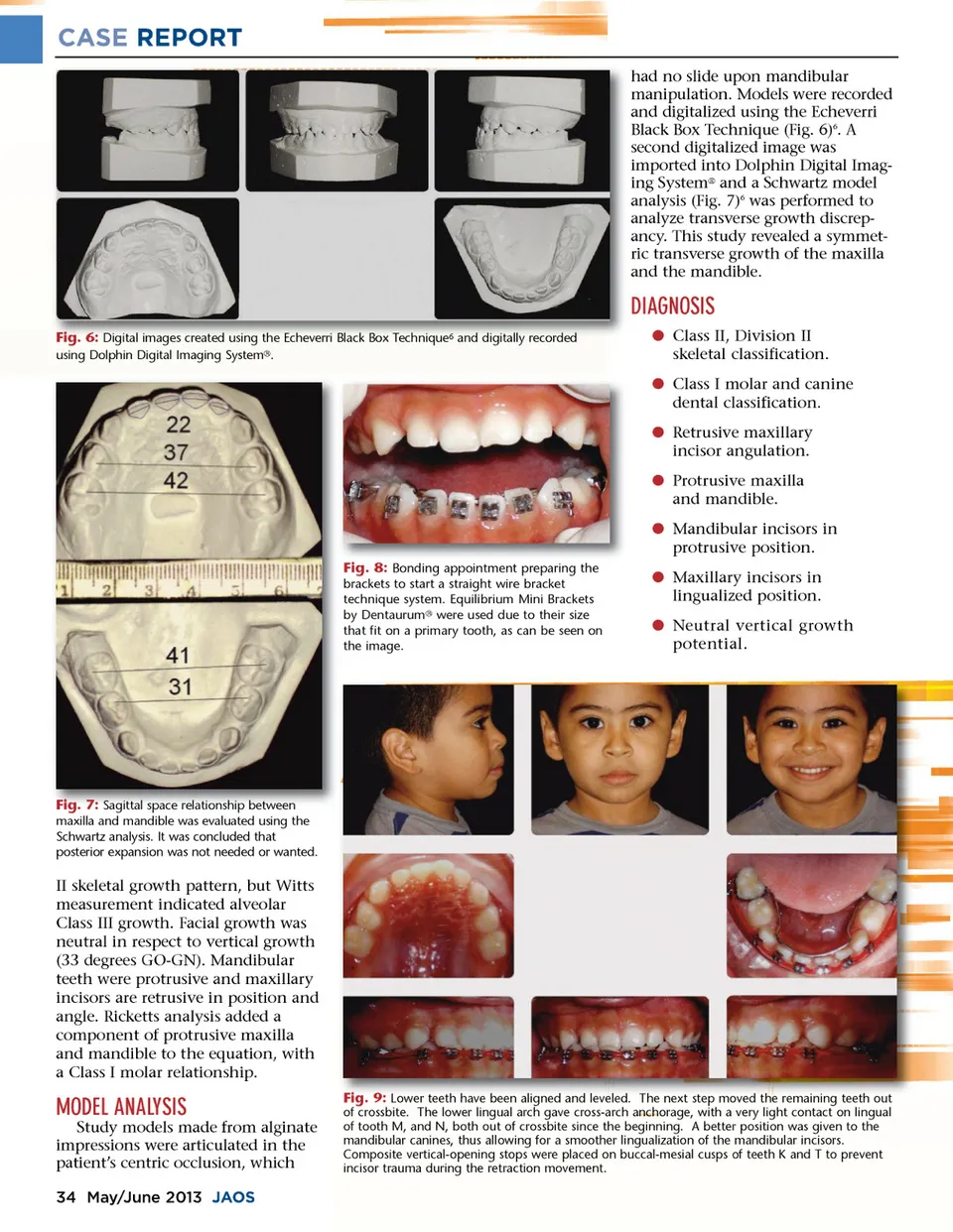
CASE REPORT had no slide upon mandibular manipulation. Models were recorded and digitalized using the Echeverri Black Box Technique (Fig. 6) 6 . A second digitalized image was imported into Dolphin Digital Imag-ing System ® and a Schwartz model analysis (Fig. 7) 6 was performed to analyze transverse growth discrep-ancy. This study revealed a symmet-ric transverse growth of the maxilla and the mandible. DIAGNOSIS Fig. 6: Digital images created using the Echeverri Black Box Technique 6 and digitally recorded using Dolphin Digital Imaging System ® . b Class II, Division II skeletal classification. b Class I molar and canine dental classification. b Retrusive maxillary incisor angulation. b Protrusive maxilla and mandible. b Mandibular incisors in protrusive position. Fig. 8: Bonding appointment preparing the brackets to start a straight wire bracket technique system. Equilibrium Mini Brackets by Dentaurum ® were used due to their size that fit on a primary tooth, as can be seen on the image. b Maxillary incisors in lingualized position. b Neutral vertical growth potential. Fig. 7: Sagittal space relationship between maxilla and mandible was evaluated using the Schwartz analysis. It was concluded that posterior expansion was not needed or wanted. II skeletal growth pattern, but Witts measurement indicated alveolar Class III growth. Facial growth was neutral in respect to vertical growth (33 degrees GO-GN). Mandibular teeth were protrusive and maxillary incisors are retrusive in position and angle. Ricketts analysis added a component of protrusive maxilla and mandible to the equation, with a Class I molar relationship. MODEL ANALYSIS Study models made from alginate impressions were articulated in the patient’s centric occlusion, which 34 May/June 2013 JAOS Fig. 9: Lower teeth have been aligned and leveled. The next step moved the remaining teeth out of crossbite. The lower lingual arch gave cross-arch anchorage, with a very light contact on lingual of tooth M, and N, both out of crossbite since the beginning. A better position was given to the mandibular canines, thus allowing for a smoother lingualization of the mandibular incisors. Composite vertical-opening stops were placed on buccal-mesial cusps of teeth K and T to prevent incisor trauma during the retraction movement.
Journal of the American Orthodontic Society May-June 2013: Page 34