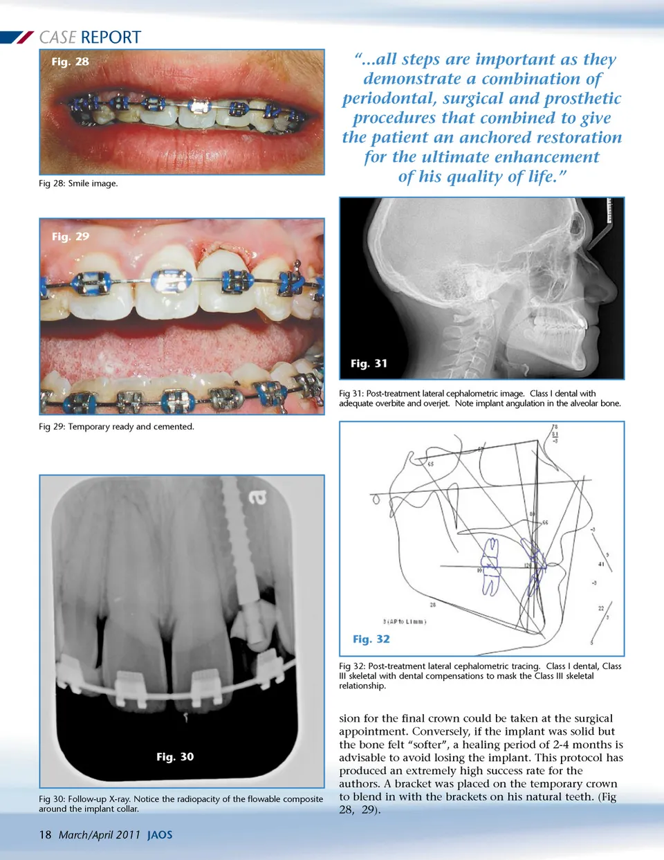
CASE REPORT Fig. 28 Fig 28: Smile image. “...all steps are important as they demonstrate a combination of periodontal, surgical and prosthetic procedures that combined to give the patient an anchored restoration for the ultimate enhancement of his quality of life.” Fig. 29 Fig. 31 Fig 31: Post-treatment lateral cephalometric image. Class I dental with adequate overbite and overjet. Note implant angulation in the alveolar bone. Fig 29: Temporary ready and cemented. Fig. 32 Fig 32: Post-treatment lateral cephalometric tracing. Class I dental, Class III skeletal with dental compensations to mask the Class III skeletal relationship. Fig. 30 Fig 30: Follow-up X-ray. Notice the radiopacity of the flowable composite around the implant collar. sion for the final crown could be taken at the surgical appointment. Conversely, if the implant was solid but the bone felt “softer”, a healing period of 2-4 months is advisable to avoid losing the implant. This protocol has produced an extremely high success rate for the authors. A bracket was placed on the temporary crown to blend in with the brackets on his natural teeth. (Fig 28, 29). 18 March/April 2011 JAOS
Journal of the American Orthodontic Society March-April 2011: Page 18